Molecular Interactions
(aka Noncovalent Interactions, Intermolecular Forces)
and the Behaviors of Biological Macromolecules
| en español | slide deck (pptx) |
Loren Dean Williams
Professor
Chemistry & Biochemistry, Georgia Tech
revised 8/17/2026
Part 1. Molecular Interactions.
A. Introduction.

Molecular interactions are attractive or repulsive forces between molecules and non-bonded atoms. They play crucial roles across chemistry, biochemistry, biophysics, and materials science, governing processes such as protein folding, drug design, pathogen detection, nanotechnology, gecko adhesion, and the origin of life. Whether referred to as noncovalent, intermolecular, or non-bonding forces, these terms all describe the same fundamental phenomena.
Overview of Non-Bonding Interactions. Molecular interactions occur between molecules and non-bonded atoms. These interactions can be cohesive (attraction between like substances), adhesive (attraction between different substances), or repulsive. During phase changes (ice melting, water boiling, carbon dioxide subliming) and biological structural transitions (protein and RNA unfolding, DNA strand separation, membrane disassembly), molecular interactions rearrange while covalent bonds remain intact. The enthalpy of a typical noncovalent interaction ranges from 1 to 10 kcal/mol (4 to 42 kJ/mol), where the lower end is on the order of thermal energy RT band the upper end remains substantially weaker than a covalent bond.
Bonding Interactions. Bonds hold atoms together within molecules. A molecule is a group of atoms that associates strongly enough that it does not dissociate or lose structure when it interacts with its environment. At room temperature two nitrogen atoms can be bonded (N2). Bonds break and form during chemical reactions. In the chemical reaction called fire, bonds of cellulose break while bonds of carbon dioxide and water form. Bond enthalpies are on the order of 100 kcal/mole (400 kjoule/mole), which is much greater than RT at room temperature.
Boiling Points. When a liquid transitions to the gas phase, intermolecular interactions are disrupted. Ideal gases are the only systems where molecular interactions are completely absent, apart from short-range repulsions during collisions. Differences in boiling temperatures provide a qualitative measure of intermolecular interaction strength in the liquid phase: higher boiling points reflect stronger attraction between molecules. For example, the boiling point of H2O is significantly higher than that of N2 because liquid water has much stronger intermolecular interactions than liquid nitrogen.
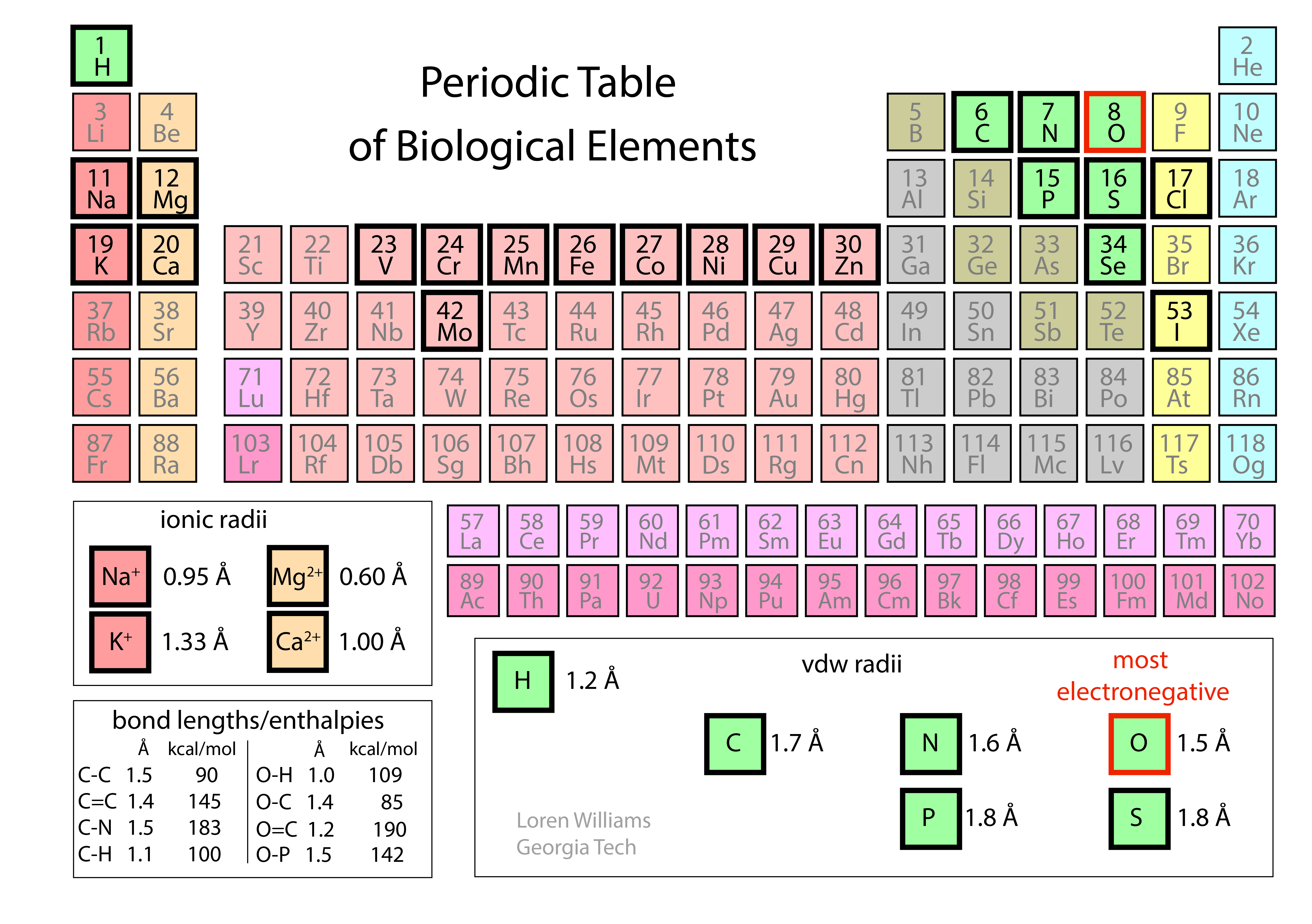
The Periodic Table. Understanding molecular interactions requires knowing molecular sizes, shapes, and electronegativities. The Periodic Table provides the foundational atomic trends that dictate these properties.
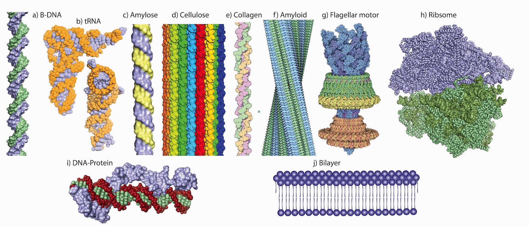
Native states. In biological systems, biopolymers assemble into defined, functional architectures known as native states. These structures can be unimolecular, such as a folded protein or tRNA, or multimolecular assemblies, such as cellulose fibrils composed of polyglucose strands or a ribosome built from multiple rRNAs and many proteins.
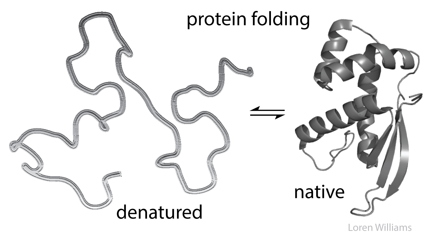
Denatured states. When a protein or RNA unfolds (denatures), DNA strands separate (melt), or a complex like the ribosome disassembles, previously buried interior regions become exposed to the aqueous surroundings. Internal interactions that stabilized the native assembly are then replaced by interactions with the surrounding water and ions.
Monster truck tug-of-war. Biological molecules are subject to powerful forces pulling in opposing directions because molecular interactions stabilize both folded and unfolded states. A vast number of intramolecular interactions within a protein in the native state are opposed by a vast number of intermolecular interactions in the denatured state, with surrounding water molecules and ions. Consequently, native macromolecules and assemblies are only marginally stable: a subtle change in pH, a slight temperature shift, or a single mutation can tip the balance and unfold a protein.
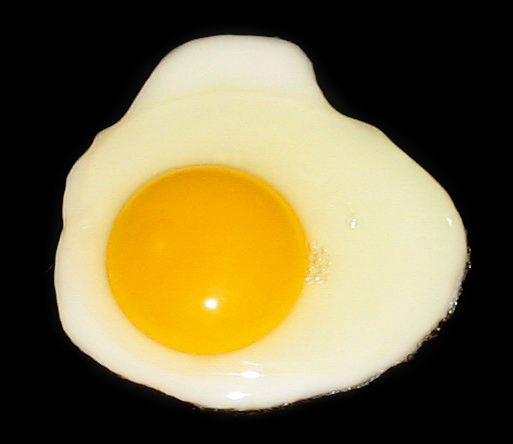
Have you ever denatured a protein (converted it from its native state to a denatured state)? Yes. When you heat an egg to around 60 °C, the albumin proteins denature and aggregate. Cooking an egg does not break covalent bonds; it changes and rearranges noncovalent molecular interactions. These unfolded proteins form large assemblies that scatter light, turning the clear egg white opaque. Similarly, adding lemon juice to milk lowers the pH, causing proteins to denature and curdle. Have you ever melted DNA? Yes, if you have ever run a PCR.
We avoid the term "van der Waals interaction." Molecular interactions were discovered by the Dutch scientist Johannes Diderik van der Waals, who observed that real gas molecules occupy finite volume and attract one another at short range, much like wet jelly beans. Unfortunately for both students and practicing scientists, the nomenclature describing these phenomena is chaotic. Terms such as noncovalent, intermolecular, non-bonding, and van der Waals are often conflated as synonyms across both "forces" and "interactions." The phrase "van der Waals interaction" has become effectively meaningless because definitions are so inconsistent and arbitrary, failing to reflect distinct physical mechanisms. Even IUPAC offers competing definitions, one of which arbitrarily excludes ion-dipole and ion-induced dipole interactions. In contrast, concepts such as "van der Waals surface" and "van der Waals radius" remain well-defined and useful (see below).
All molecular interactions are fundamentally electrostatic in nature and can be described by some variation of Coulombs Law. However, we reserve the term 'electrostatic interaction' to describe interactions between charged species (ions). Interactions between partial charges are given other names.
Molecular interactions can be classified in various ways. The categories used here (see Table of Contents) provide a clear, accessible framework aligned with standard usage across the scientific literature.
Lennard-Jones. The Lennard-Jones potential is an empirical model used to describe specific molecular interactions. However, it does not account for all noncovalent forces; formal electrostatic interactions and fixed-dipole alignments are explicitly excluded from the model.
B. Short range repulsion.
Atoms take space. Force two atoms together and they will push back. As two atoms approach one another, their occupied electron clouds begin to overlap, driving strong repulsion. This repulsive force operates over very short distances, rising steeply as interatomic separation decreases. The allowed distance between non-bonded atoms depends on atomic identity and, for metals, the ionization state as shown in the Periodic Table.
The repulsive energy scales as (di/R)12, where R is the distance between two atoms and di is the distance threshold below which repulsion dominates. This threshold, di, is determined by the van der Waals radii of the interacting atoms. The large exponent means that once R < di, even small decreases in distance trigger steep increases in repulsive energy. Short-range repulsion only becomes significant at close proximity (R < di), but at these contact distances, it overwhelms all other forces. Because this repulsion rises so sharply as distance decreases, it is often useful to treat atoms as hard spheres (like miniature billiard balls) with defined boundaries (van der Waals surfaces) and fixed radii (van der Waals radii).
As two atoms approach each other, their van der Waals surfaces make contact when the distance between them equals the sum of their van der Waals radii. Pushing them any closer causes repulsive energy to skyrocket, making this sum the practical limit for how close two non-bonded atoms can get. For example, a sulfur atom and a carbon atom can come no closer together than:
rS + rC = 1.8 Å + 1.7 Å = 3.5 Å
This assumes, of course, that no chemical bond forms. When two atoms form a covalent bond, their nuclei draw much closer together, causing violoation of van der Waals surfaces.
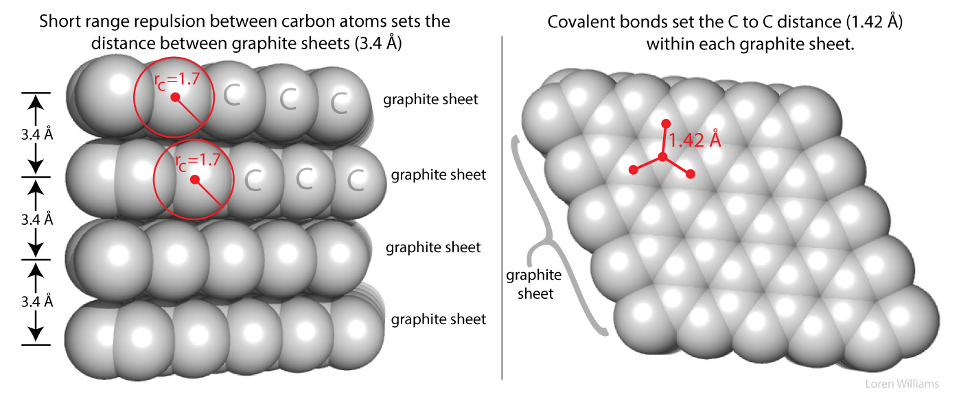
Short-range repulsion is part of your everyday experience. It prevents your hands from passing through each other when you clap, and it keeps ordinary matter from collapsing into ultra-dense states (~1014 g/mL) typical of atomic nuclei. Immense gravitational forces, such as those in neutron stars, can overcome short-range repulsion and collapse atomic structures.
Here on Earth, with our modest gravity, the van der Waals radius of carbon (rC) is illustrated by the spacing between graphite sheets. The distance between carbon atoms in adjacent layers is roughly twice the van der Waals radius of carbon (2 × rC = 2 × 1.7 Å = 3.4 Å). In contrast, carbon atoms within a single sheet are covalently bonded, causing their electron clouds to interpenetrate: these in-plane atoms are separated by only 1.42 Å, far shorter than the non-bonded contact distance. As discussed in other sections, van der Waals surfaces also interpenetrate during hydrogen bonding. Atomic coordinates for graphite are available here [coordinates].
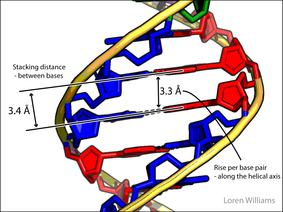
In both DNA and RNA duplexes, the distance between adjacent stacked base pairs is 3.4 Å, identical to the spacing between sheets of graphite. In a B-form helix (DNA), base pairs are only slightly inclined, with their normals nearly parallel to the helical axis; therefore, the rise per base pair along the axis is only slightly less than the 3.4 Å stacking distance. In an A-form helix (RNA), base-pair inclination is much greater, reducing the axial rise per base pair to approximately 2.3 Å.
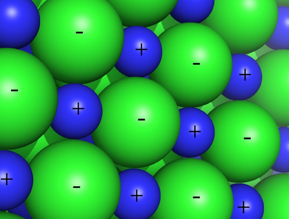
How do you sense short range repulsion? Try compressing a liquid.
C. Electrostatic interactions.
Electrostatic interactions occur between cations and anions, species bearing net integer charges (… -2, -1, +1, +2, …). These forces can be either attractive or repulsive depending on the signs of the charges: like charges repel, while opposite charges attract.
Strong attractive electrostatic forces give sodium chloride and other salts exceptionally low vapor pressures. If you place crystals of table salt (NaCl, composed of Na+ cations and Cl- anions) in a hot pan, they will not vaporize or sublime because the lattice interactions holding the crystal together (ionic bonds) are extraordinarily robust. When isolated cations and anions interact within a folded protein or RNA (often called salt bridges or ion pairs), they are classified as noncovalent electrostatic interactions. These forces are strong and operate over long ranges: electrostatic force fall of gradually with distance, as 1/r2 (with potential energy scaling as 1/r, where r is the inter-ion distance).
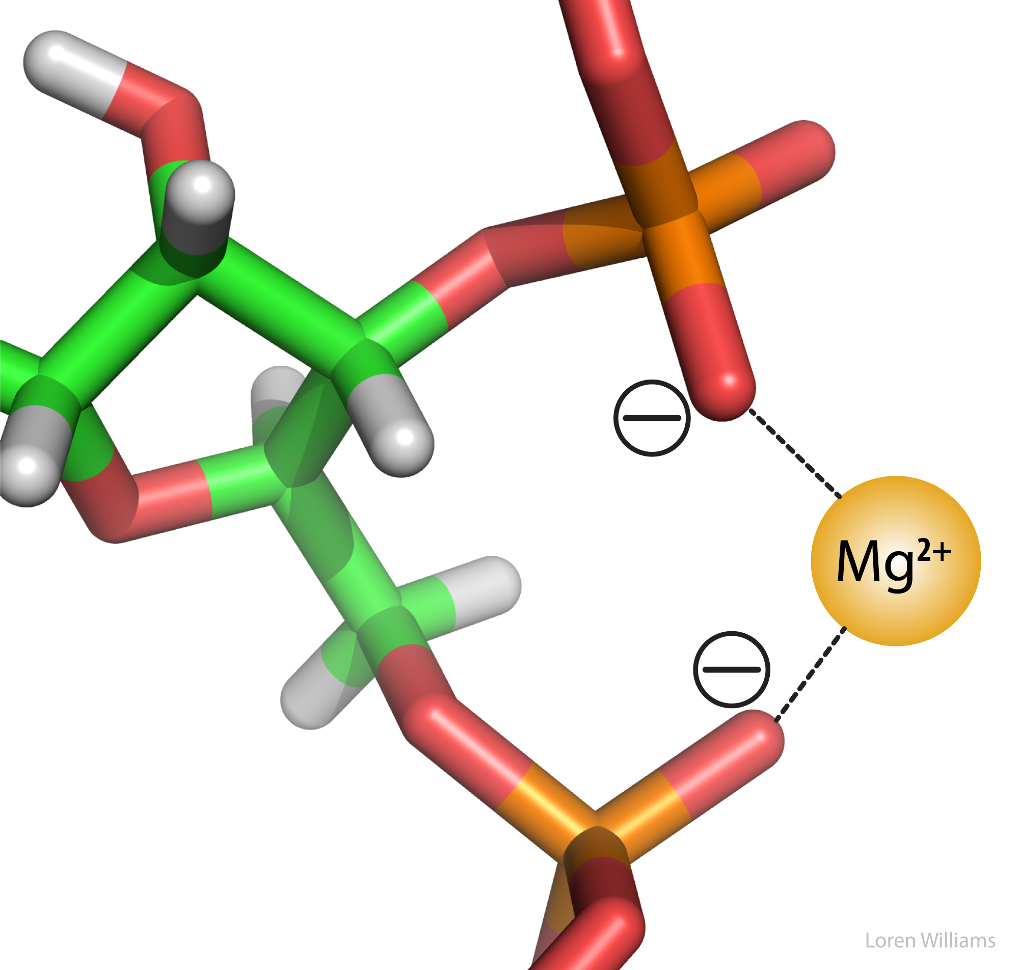
Electrostatic attraction stabilizes the association between the negatively charged phosphate oxygens of RNA (formal charge of -1 per phosphate) and divalent magnesium ions (Mg2+, charge = +2), as shown in the figure below. Magnesium ions serve as crucial counterions associated with both RNA and DNA in vivo, while calcium ions (Ca2+, charge = +2) associate with many regulatory and structural proteins. In protein and RNA folding, as well as DNA duplex annealing, electrostatic forces depend strongly on both salt concentration and pH. As discussed later in this document, electrostatic interactions are attenuated (dielectrically screened) by water.
Favorable electrostatic interactions between paired anionic and cationic amino acid side chains are common in folded proteins. Ion pairs, often called salt bridges, form when the charged group of a cationic amino acid (such as lysine or arginine) comes within roughly 3.0 to 5.0 Å of an anionic amino acid (such as aspartate or glutamate). At these contact distances, the charged functional groups typically engage in hydrogen bonding in addition to electrostatic interactions.
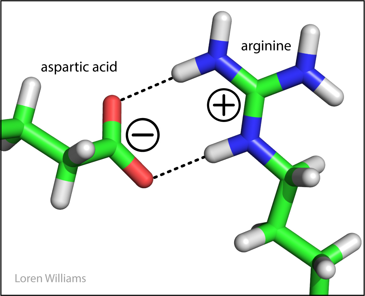
The electrostatic force between two point charges is given by Coulomb's Law:
Force = k q1 q2 / (ε r2)
where k = 9.0 × 10⁹ N·m2 / C2
q1, q2 = charges of the interacting species (unit of charge = 1.6 × 10⁻¹⁹ C for a single electron or proton)
r = distance between the point charges (meters)
ε = dielectric constant of the medium (unitless)
The dielectric constant, ε, reflects the ability of a medium to shield (screen) interacting charges from one another. ε is defined as 1 in a vacuum, roughly 4 in the interior of a protein, and approximately 80 in bulk water. Because water is so effective at shielding charges, it dramatically attenuates electrostatic forces between ions. Calculating electrostatic effects in biological systems is exceptionally complex because the dielectric medium is highly non-uniform: dielectric microenvironments vary widely across a macromolecule, with minimal charge shielding in regions dominated by hydrocarbon amino acid side chains and substantial shielding in regions rich in polar side chains or bulk water.
The electrostatic potential energy per mole is given by:
ΔE = k a q1 q2 / (ε r)
where a = Avogadro's number (yielding energy per mole).
One can roughly estimate the energetics of a charge-charge interaction within a biopolymer. The electrostatic energy between a protonated amino group (charge +1) and a carboxylate group (charge -1) separated by 4 Å in the low-dielectric interior of a protein (ε = 4) is given by:
ΔE = -(9.0 × 10⁹ N·m2/C2)(6.02 × 1023 mol-1)(1.6 × 10⁻¹⁹ C)2 / [4 × (4 × 10-10 m)]
= -87 kJ/mol = -21 kcal/mol
This rough approximation is nearly an order of magnitude larger than values determined experimentally. In practice, an ion pair contributes a favorable ΔG of only 1 to 4 kcal/mol (4.2 to 16.7 kJ/mol) to the net stability of a folded protein. The calculated electrostatic gain is partially offset by the desolvation penalty, which requires shedding favorable interactions between the charged side chains and water molecules.
A note on nomenclature. The attractive forces between a Mg2+ ion and nucleic acid phosphate groups are classified as electrostatic interactions because both partners carry net integer charges (… -2, -1, +1, +2, …). Terms such as dipole-dipole, dipole-induced dipole, and London dispersion are used to describe interactions involving partial charges. This naming convention can be confusing because, at a fundamental physical level, all molecular interactions are electrostatic in origin: they arise entirely from Coulombic forces between electrons and atomic nuclei. Nevertheless, by standard chemical convention, the term "electrostatic interaction" is typically reserved for interactions between species bearing full integer charges.
D. Dipolar Interactions.
| Atom | Electronegativity (unitless) |
|---|---|
| H | 2.2 |
| C | 2.6 |
| N | 3.0 |
| O | 3.5 |
| P | 2.2 |
| S | 2.5 |
Electronegativity. Before you can understand dipolar interactions, you have to understand electronegativity. In chemical bonds between different elements, shared electrons are not distributed equally. The intrinsic ability of an atom within a molecule to pull bonding electron density toward itself is defined as its electronegativity. Fluorine is the most electronegative element (4.0), while cesium is the least electronegative (0.7). Electronegativity increases from left to right across a period as effective nuclear charge increases, and it increases from bottom to top up a group as atomic radius decreases and bonding electrons experience less core electron shielding.
Partial Charges. In bonds between atoms of unequal electronegativities, the bonding electron cloud is displaced toward the more electronegative partner. The atom with lower electronegativity carries a partial positive charge (δ+), while the more electronegative atom carries a partial negative charge (δ-). A greater difference in electronegativity between bonded atoms produces a more polar bond with partial charges of larger magnitude. In biological systems, oxygen is the most electronegative element (followed by nitrogen), regularly bearing the largest partial negative charge in biomolecules.
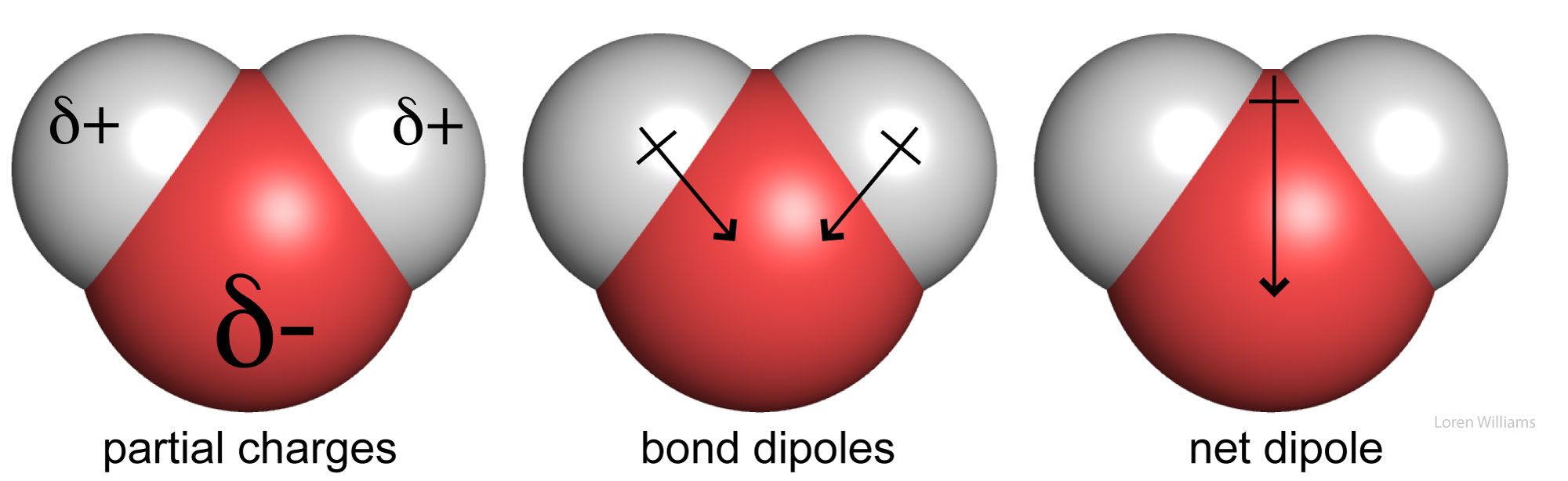
In methanol (CH3OH) and water (H2O), the electronegative oxygen atom draws electron density away from bonded carbon and hydrogen atoms, leaving a partial negative charge on oxygen and partial positive charges on the carbon and hydrogens. This phenomenon of internal charge separation defines molecular polarity. Both methanol and water are polar molecules. In contrast, molecular nitrogen (N2) is nonpolar because the two nitrogen atoms possess identical electronegativities and share electrons equally. Similarly, hydrocarbons (such as alkanes, CH3CH2…CH2CH3) are nonpolar because the electronegativities of carbon (2.55) and hydrogen (2.20) are very similar, resulting in an essentially uniform distribution of charge.
Dipole Moment. The extent of charge separation within a bond or molecule is quantified by its dipole moment, μ. The magnitude of a dipole moment is determined by the product of the separated partial charges and the distance between them. By chemical convention, dipole moments are typically expressed in electrostatic units (esu) for charge and centimeters for distance, which define the Debye unit (1 D = 10-18 esu·cm). The dipole moment of a full elementary charge separated by 1 Å equals:
(4.8 × 10⁻¹⁰ esu) × (1.0 × 10⁻⁸ cm) = 4.8 × 10⁻¹⁸ esu·cm
= 4.8 Debye (4.8 D)
For comparison, the net dipole moment of water is 1.85 D (HCl = 1.1 D; CH3Cl = 1.9 D; HCN = 2.9 D; NH3 = 1.47 D).
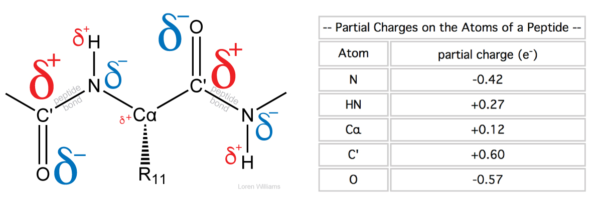
Resonance within the planar peptide unit (amide group) contributes substantial partial charges to the backbone atoms: the carbonyl oxygen carries a partial negative charge of roughly δ- ≈ -0.4 to -0.5 e, balanced by a partial positive charge on the carbonyl carbon (δ+ ≈ +0.4 to +0.5 e). On the other side of the bond, the amide hydrogen bears a partial positive charge of roughly δ+ ≈ +0.3 e relative to the amide nitrogen. Together, this charge distribution generates a large net dipole moment of approximately 3.7 Debye (3.7 D) that aligns roughly parallel to the N-H and C=O bonds, directed from the partial positive amide group toward the partial negative carbonyl oxygen.
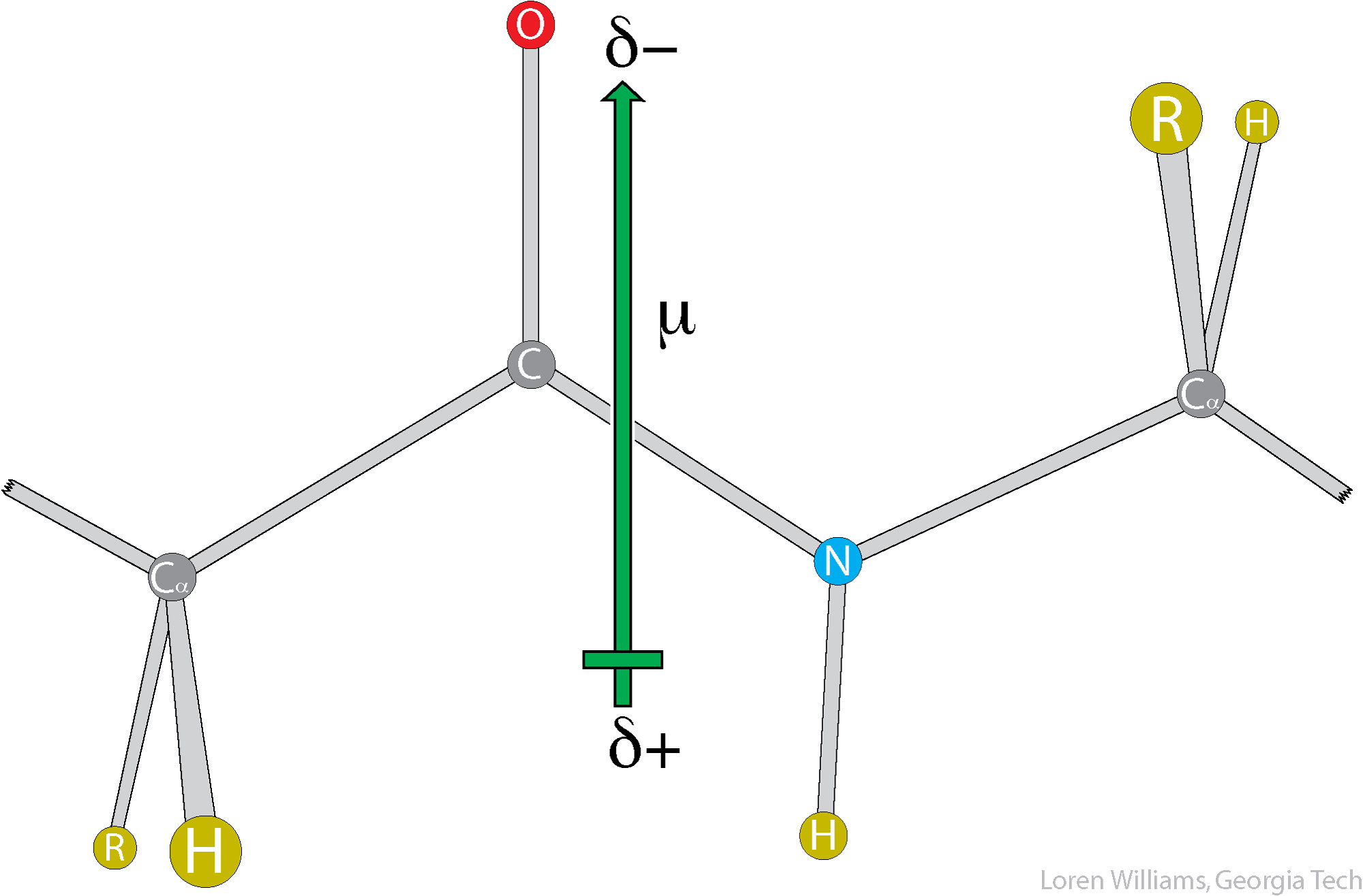
Given the substantial dipole moment of each peptide bond, one would expect dipolar interactions to play a central role in protein folding, secondary structure stability, and macromolecular recognition. They do.
A permanent dipole is surrounded by an electric field that exerts electrostatic forces on neighboring charged and partially charged species. Interactions between dipoles and ions are termed ion-dipole (or charge-dipole) interactions. Dipoles also interact directly with other permanent dipoles (dipole-dipole interactions) and can polarize the electron clouds of surrounding molecules (dipole-induced dipole interactions). Each of these interaction types is discussed separately in the sections below.
Dipole–dipole interactions arise from electrostatic forces between permanent molecular dipoles. The strength of the interaction depends on dipole magnitudes, separation distance, and relative orientation. Colinear head-to-tail orientations and antiparallel side-by-side arrangements are attractive, whereas head-to-head and parallel side-by-side configurations are repulsive. The values below reflect interaction energies for two 1 D dipoles separated by 5 Å in a dielectric medium of ε = 4.
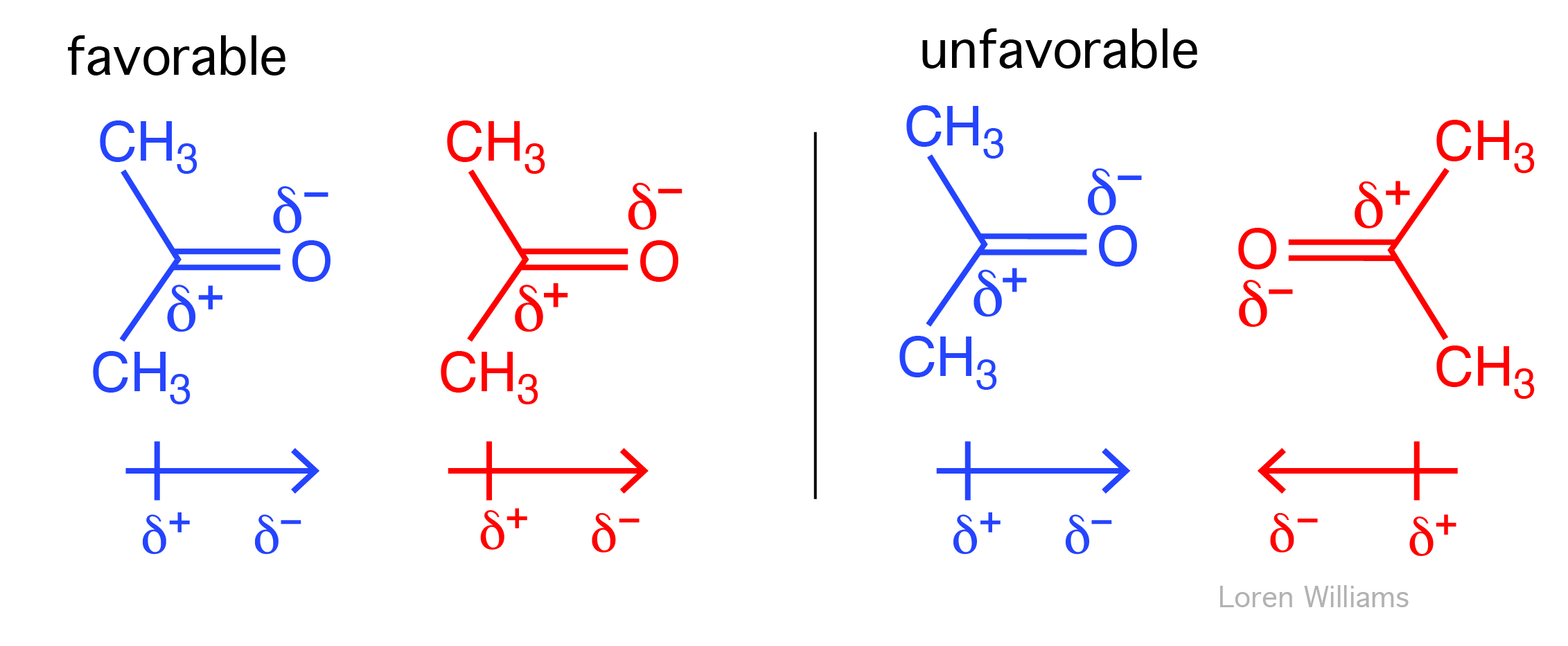
In liquids, rapid thermal tumbling continuously alters molecular orientations. Because lower-energy, attractive configurations are statistically favored, net dipole–dipole interactions remain attractive (as in liquid acetone). While fixed dipole–dipole interactions scale with 1/r3, thermal rotational averaging in liquid media causes the net attraction to scale with 1/r6.

Like a windsock shifting with the wind, the electron cloud of a polarizable molecule is continuously distorted by the electric fields of tumbling polar neighbors. This transient polarization induces a dipole that aligns favorably with the surrounding permanent dipoles, generating a net attractive interaction (1/r6).
The electric field surrounding an ion or permanent dipole distorts the electron distribution of adjacent molecules. This distortion is called polarization. A molecule's susceptibility to such deformation is its polarizability (α). Large, diffuse electron clouds are the most polarizable: xenon is more polarizable than helium, and aromatic amino acid side chains with delocalized π electrons (phenylalanine, tryptophan) are significantly polarizable than aliphatic side chains (isoleucine).
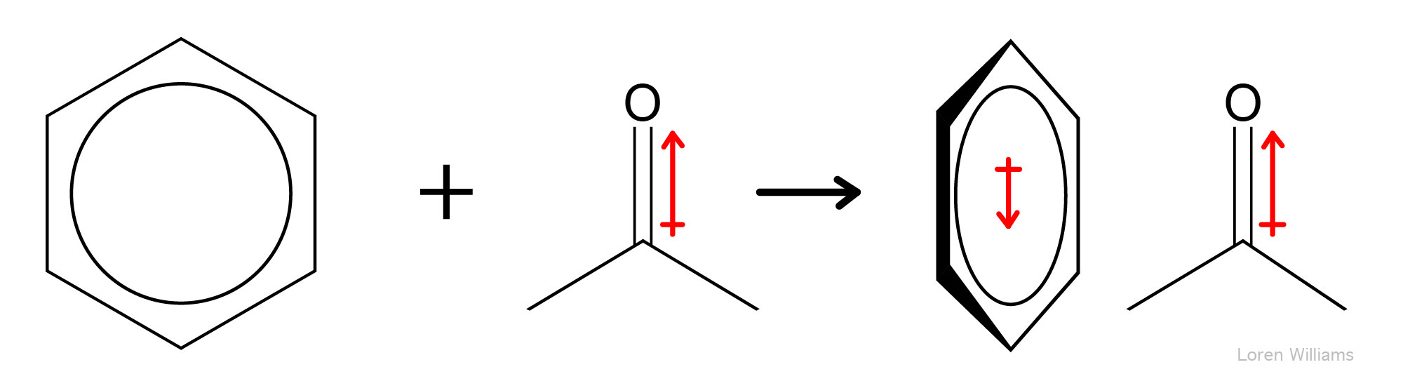
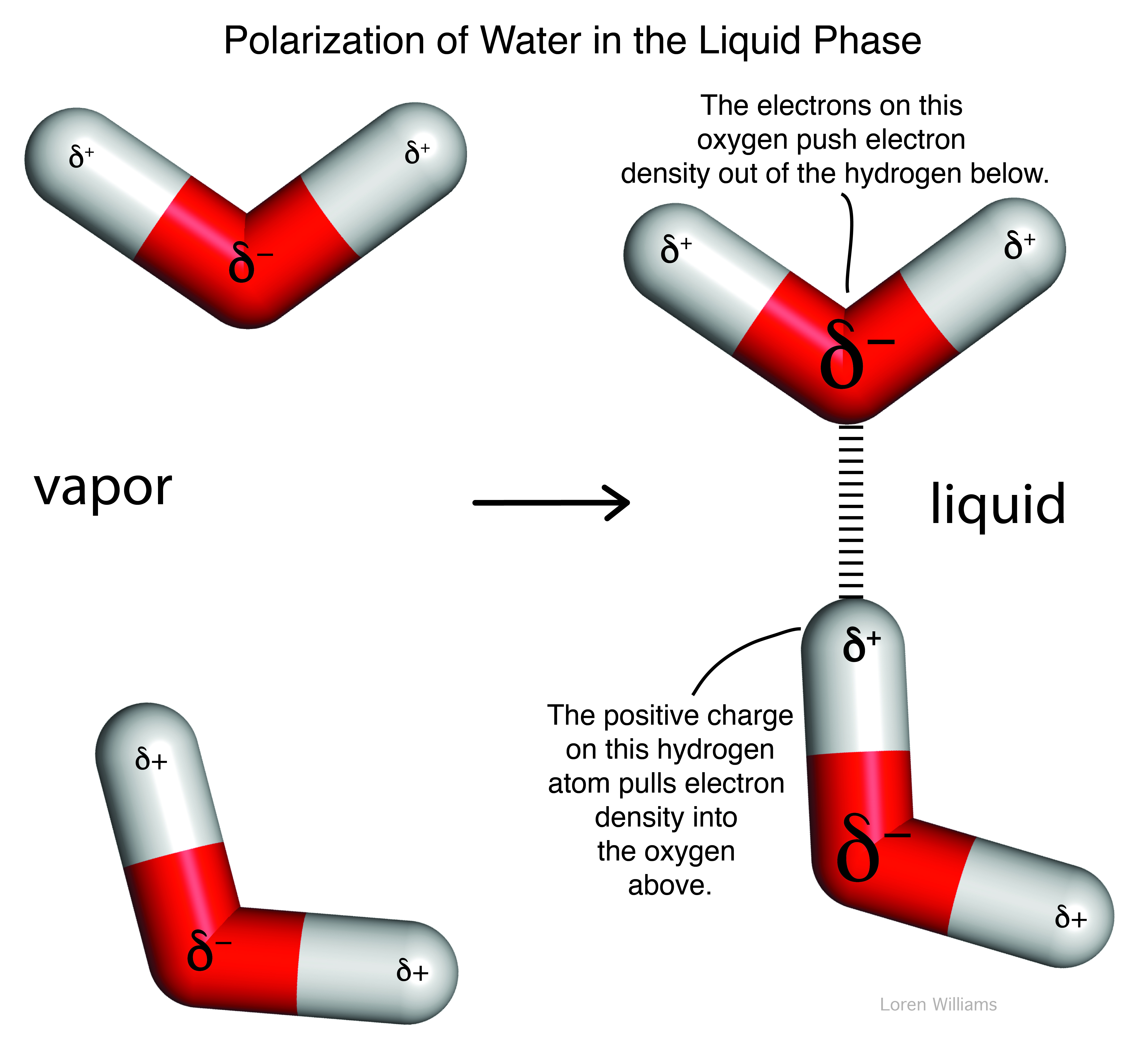
Dipole–induced dipole interactions operate even among molecules possessing permanent dipoles. In polar liquids such as water, neighboring molecules mutually polarize one another: the electric field of each water molecule continually perturbs and enhances the dipole moments of surrounding water molecules.
Dipole–induced dipole interactions are always attractive, contribute up to ~0.5 kcal/mol (2.1 kJ/mol) toward molecular association, and scale with 1/r6. Species with interger charge (e.g., Na+, Mg2+, −COO−) generate strong electric fields, producing charge–induced dipole (or ion–induced dipole) interactions that scale with 1/r4 and play critical stabilizing roles in proteins and nucleic acids.
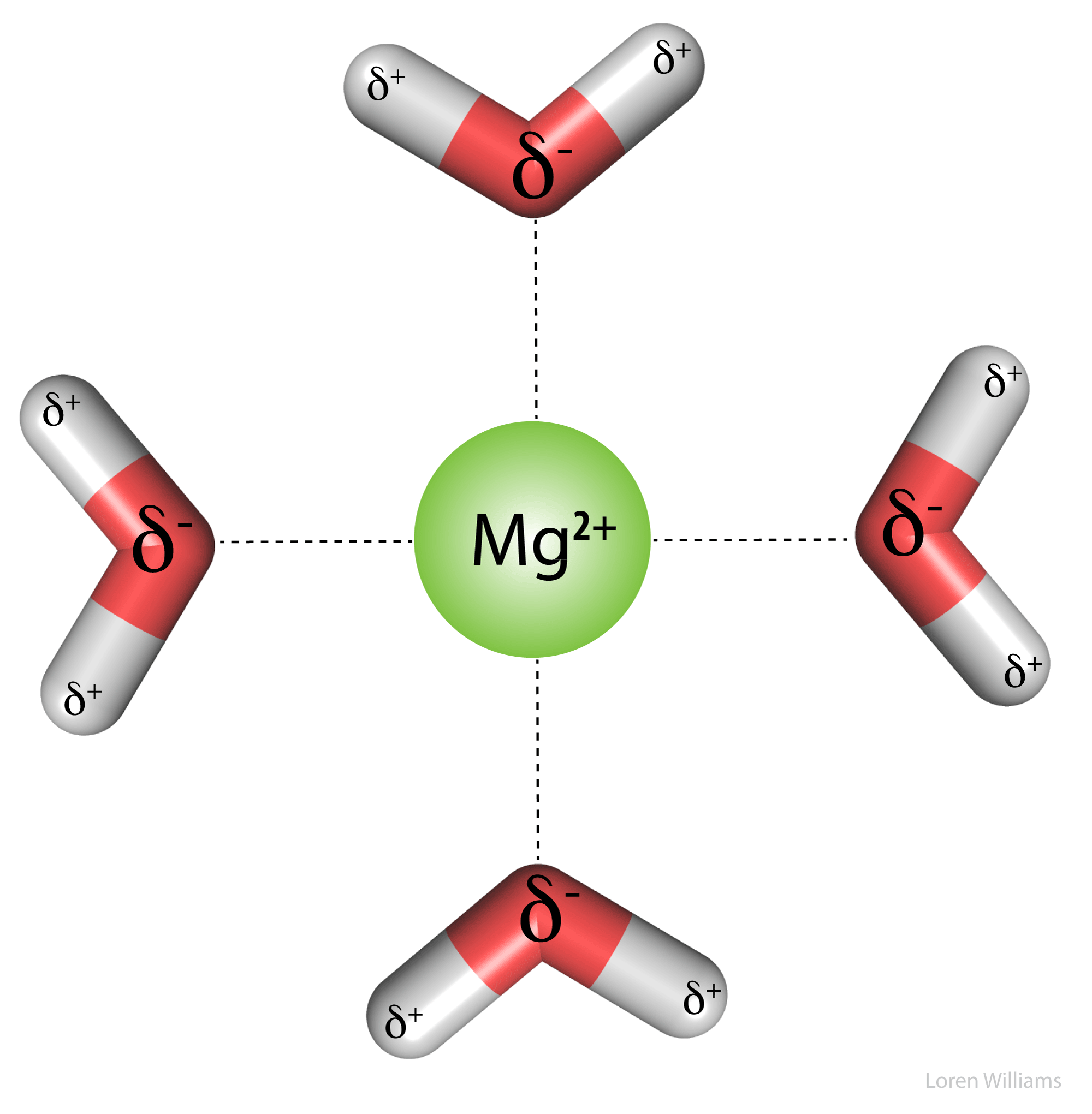
Charge–dipole (or ion–dipole) interactions arise from electrostatic forces between ions and permanent dipoles, scaling with 1/r2. These interactions drive the high aqueous solubility of salts in water. Water's strong dipole interacts favorably with both Na+ cations and Cl− anions to form stabilizing hydration shells.
E. Fluctuating dipolar interactions (Dispersive interactions, London Forces).
Resonance occurs across diverse physical systems: a child pumping a swing, tidal oscillations in the Bay of Fundy, and the harmonic vibrations of violin strings all demonstrate resonance. The collapse of the Tacoma Narrows Bridge remains an iconic macroscopic demonstration of resonance.
Resonance also emerges from collective human motion. During a 2023 concert in Seattle, 70,000 fans dancing in phase to "Shake It Off" generated localized ground acceleration equivalent to a magnitude 2.3 earthquake. Similarly, when London's Millennium Bridge opened in 2000, subtle lateral sways prompted pedestrians to unconsciously synchronize their strides with the deck's motion. This positive feedback loop drove violent side-to-side oscillations. Retrofitted dampers were installed to dissipate the vibrational energy.
Atoms and molecules exhibit electronic resonance. Although noble gases like helium and xenon possess spherically symmetric electron clouds on average, their electron densities fluctuate at characteristic quantum frequencies. An instantaneous snapshot of an atom reveals a transient, asymmetric charge distribution—an instantaneous dipole—even though its time-averaged state is non-polar and spherical.
Because electron distributions fluctuate continuously, so do dipole moments. In condensed phases (liquids and solids), adjacent fluctuating dipoles couple electrostatically. Driven by electron–electron repulsion, electron motions across neighboring molecules become correlated, oscillating in phase like coupled resonators. This synchronized alignment produces a net, universally attractive force termed London dispersion (scaling with 1/r6).
This behavior is like two people jumping on a trampoline: the flexible mat transfers energy between them, coupling their motions. Jumping out of sync creates jarring resistance, naturally driving their bounces into a coordinated rhythm. In condensed phases, the electrostatic field acts like the trampoline mat, coupling neighboring atoms so their fluctuating electron clouds spontaneously correlate into an attractive, synchronized motion.
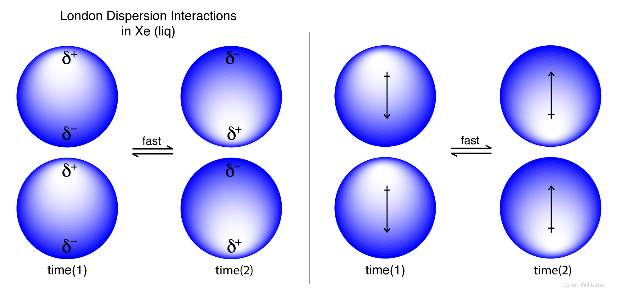
London dispersion interactions are universally attractive, operating between all adjacent atoms and molecules regardless of static dipoles. Their magnitude scales with polarizability (α), as illustrated by the increase in noble gas boiling points: He (4.2 K), Ne (27 K), Ar (87 K), Kr (120 K), and Xe (165 K). Because noble gases lack permanent dipoles, dispersion is the only cohesive force enabling their liquid and solid states. Dispersion forces are particularly strong in aromatic systems, whose delocalized π electron clouds are very polarizable.
Within the densely packed interior of a folded protein, the vast number of pairwise atom–atom contacts makes London dispersion a dominant contributor to overall stability. Because dispersion scales directly with polarizability, aromatic side chains (tryptophan, tyrosine, phenylalanine, and histidine) engage in the strongest dispersive interactions, driving the tight packing of hydrophobic cores and stability of native folds.
What about water? Even polar molecules capable of strong hydrogen bonding experience significant dispersion. In liquid water, London dispersion forces account for approximately 25% of the total intermolecular attractive energy.
F. Cation-Π interactions.
| Cation-Π interactions with benzene (gas phase) |
|
|---|---|
| Ion | ΔH (kcal/mol) |
| Li+ | -38 |
| Na+ | -28 |
| K+ | -19 |
| NH4+ | -19 |
The π-systems of aromatic rings (such as benzene, tryptophan, tyrosine, and phenylalanine) concentrate partial negative charge above and below the ring plane. A cation can interact favorably with this electron-rich face, adopting a geometry centered over the ring in direct van der Waals contact. As shown in the table above, gas-phase cation–π interaction enthalpies are comparable in magnitude to cation hydration enthalpies, making cation–π interactions roughly similar in strength to aqueous ion–dipole interactions. Complex stability increases with higher cation charge density, while electron-donating and electron-withdrawing ring substituents systematically strengthen or weaken the interaction.
Cation–π interactions are important in protein architecture, where the aromAatic guanidinium group of arginine and the ε-ammonium (ε-NH3+) of lysine pack against aromatic side chains. A favorable cation–π pair contributes stabilizing energy comparable to a typical hydrogen bond or salt bridge (~2–5 kcal/mol). Tryptophan is the most prevalent π-partner and arginine the most frequent cation.
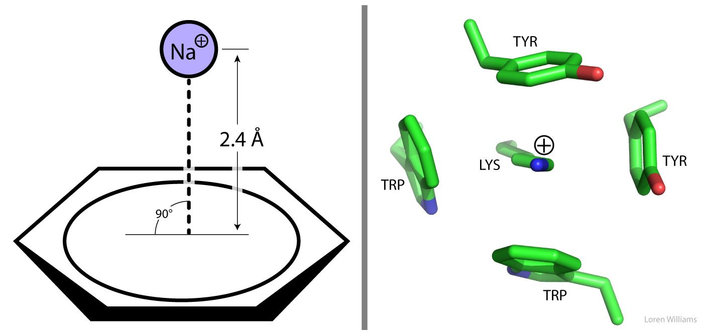
Figure 17. Left: Optimal geometry for a cation–π interaction between Na+ and benzene. The distance from the Na+ cation to the aromatic ring centroid is 2.4 Å (ionic radius of Na+ = 0.9 Å; van der Waals radius of carbon, rC = 1.7 Å). Right: The ε-ammonium group (ε-NH3+) of a lysine residue coordinated in an "aromatic box" by two tryptophan and two tyrosine side chains in glucoamylase (PDB ID: 1GAI).
G. Hydrogen Bonding.
The concept that a single hydrogen atom could act as a bridge between two electronegative atoms was formally proposed in 1920 by Wendell Latimer and Worth Rodebush in the laboratory of G.N. Lewis. Maurice Huggins, also a student in Lewis's group, independently described the hydrogen bond in his 1919 thesis.
A hydrogen bond is an attractive interaction between an electron pair on a Lewis base (the hydrogen bond acceptor, A) and a hydrogen atom partially deshielded by covalent attachment to an electronegative atom (the hydrogen bond donor, D, typically N, O, or S). In typical D–H···A architecture, the exposed hydrogen bridges the two electronegative centers, stabilized by a primarily by electrostatic attraction, but also by aligned dipoles, and partial covalent (charge-transfer) character.
Why hydrogen? Hydrogen is special because it possesses no inner core electrons. Its single electron resides in the 1s orbital and participates directly in the covalent σ bond to an electronegative atom (N, O, or S). This bonding electron density is polarized toward the electronegative partner. The hydrogen nucleus, a proton, is virtually unshielded on its distal side. This unshielded positive charge permits an unusually close approach and strong electrostatic interaction with an acceptor lone pair. In all heavier elements, filled inner electron shells shield the nucleus, preventing direct electrostatic exposure.
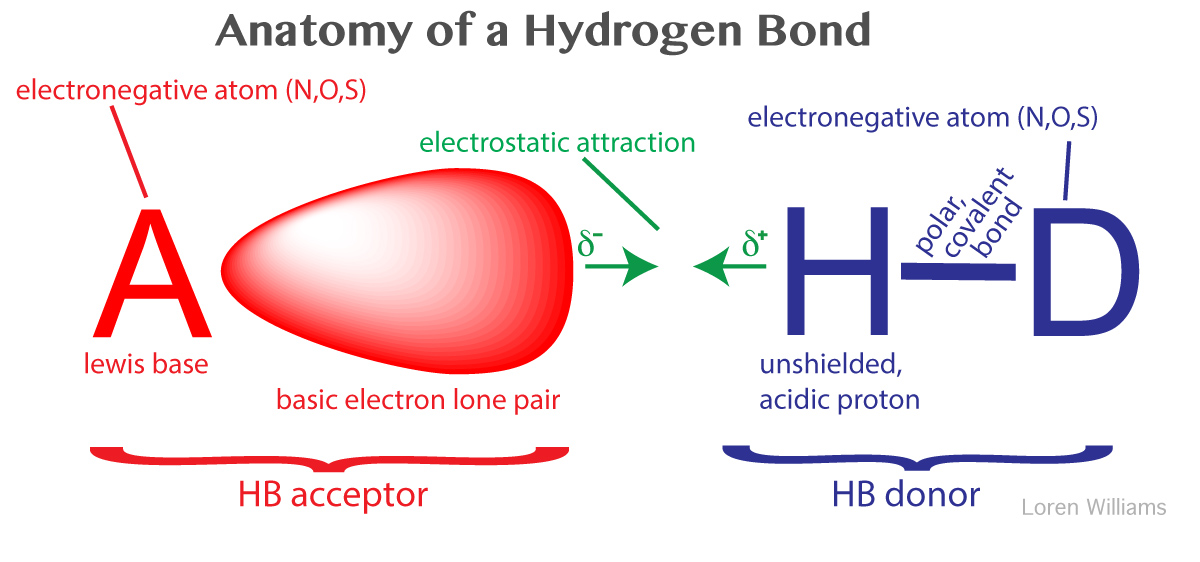
A hydrogen bond is not a Brønsted acid–base reaction where the proton is fully transferred to generate D− and HA+ ions. In a standard hydrogen bond, the proton remains covalently attached to the donor atom (D) while interacting non-covalently with the acceptor (A). Nevertheless, hydrogen bond strength correlates directly with donor acidity and acceptor basicity: more acidic donors (lower pKa) and more basic acceptors (higher pKa of the conjugate acid) form stronger hydrogen bonds, reflecting an incipient but incomplete proton transfer along the reaction coordinate.
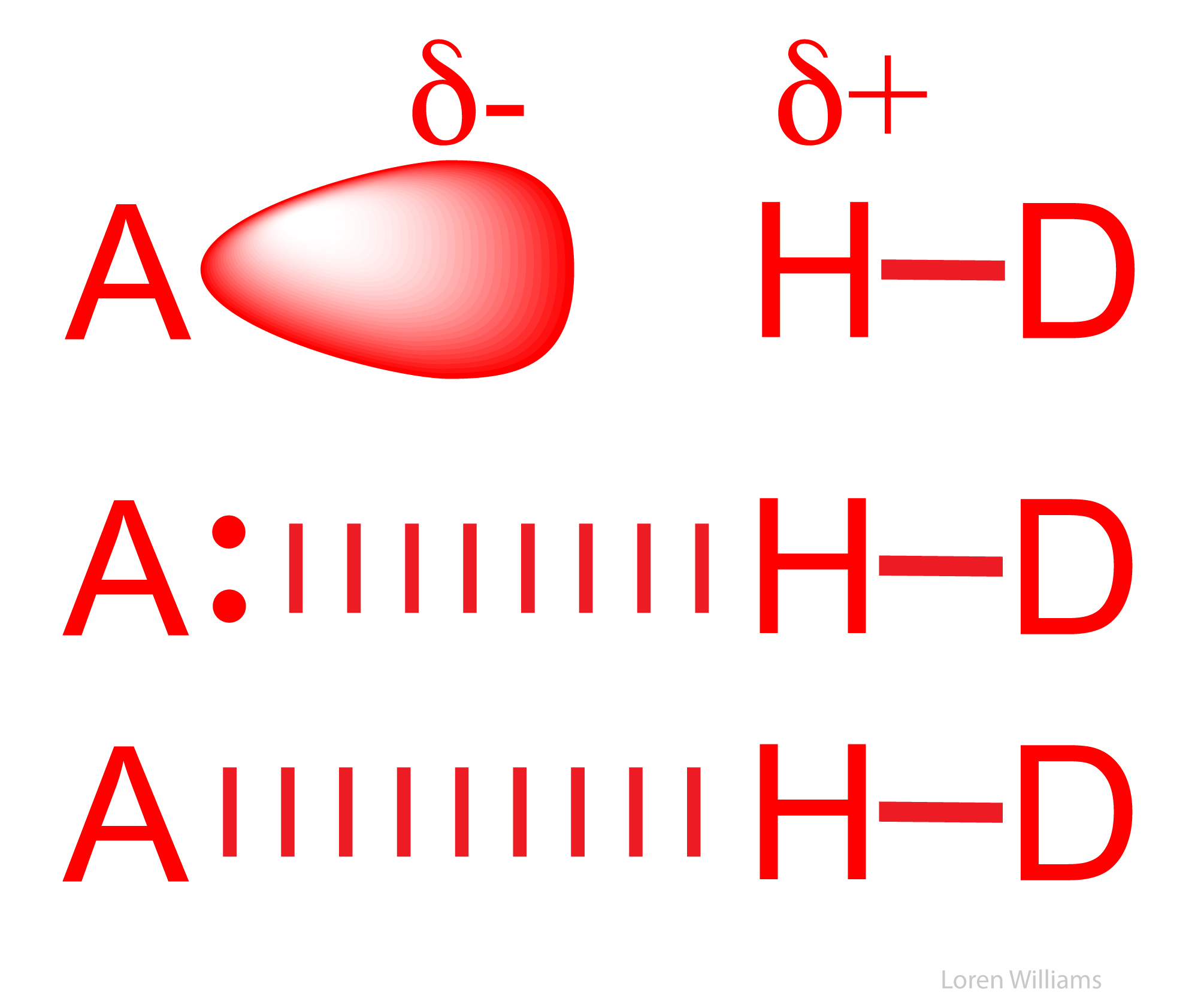
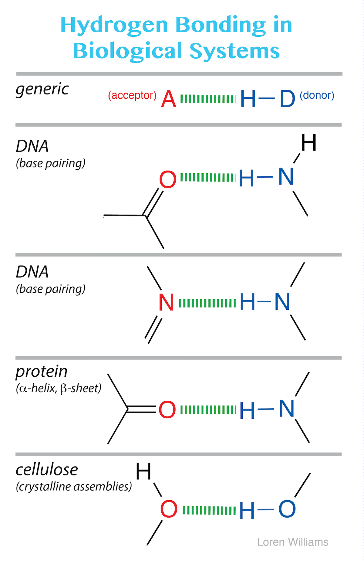
The most common hydrogen bonds in biological systems involve oxygen and nitrogen atoms as A and D. Carbonyl oxygens (C=O), ring nitrogens, and amine lone pairs (−NH2) act as the primary acceptors in nucleic acids and proteins, while amines and amide groups (−NH−) serve as the principal donors. Hydrogen bonds in glycans involve hydroxyl groups (−OH) as both donors and acceptors.
Across the periodic table, increasing the electronegativity of the donor atom (D) polarizes the D–H bond more strongly, enhancing the partial positive charge on hydrogen and strengthening the resulting hydrogen bond. While thiols (−SH) can act as both donors and acceptors, their interactions are comparatively weak due to sulfur's lower electronegativity. In contrast, carbon does not participate in classical hydrogen bonding: carbon's low electronegativity cannot sufficiently polarize C–H bonds to serve as a donor, and neutral carbon lacks the lone pairs required to act as an acceptor.
Hydrogen bond strengths span a broad continuum. Strong hydrogen bonds (20–40 kcal/mol / 80–170 kJ/mol), typically formed between charged donors and acceptors, approach the strength of covalent bonds. Moderate hydrogen bonds (3–12 kcal/mol / 12–50 kJ/mol) represent the most common category in biological macromolecules, occurring between neutral donors and acceptors. Weak hydrogen bonds (1–3 kcal/mol / 4–20 kJ/mol), such as polarized C–H···O contacts, are energetic equivalents of standard dipole–dipole interactions.
A note on nomenclature: In spite of its name, a hydrogen bond is not a bond; it is a non-covalent, non-bonding, intermolecular interaction. Covalent bonds involve shared electron pairs that establish molecular connectivity (typically 50–100 kcal/mol). Hydrogen bonds are predominantly electrostatic attractions that are an order of magnitude weaker (2–10 kcal/mol). Although the historical term hydrogen bond will persist, do not confuse hydrogen bonds with true chemical bonds.
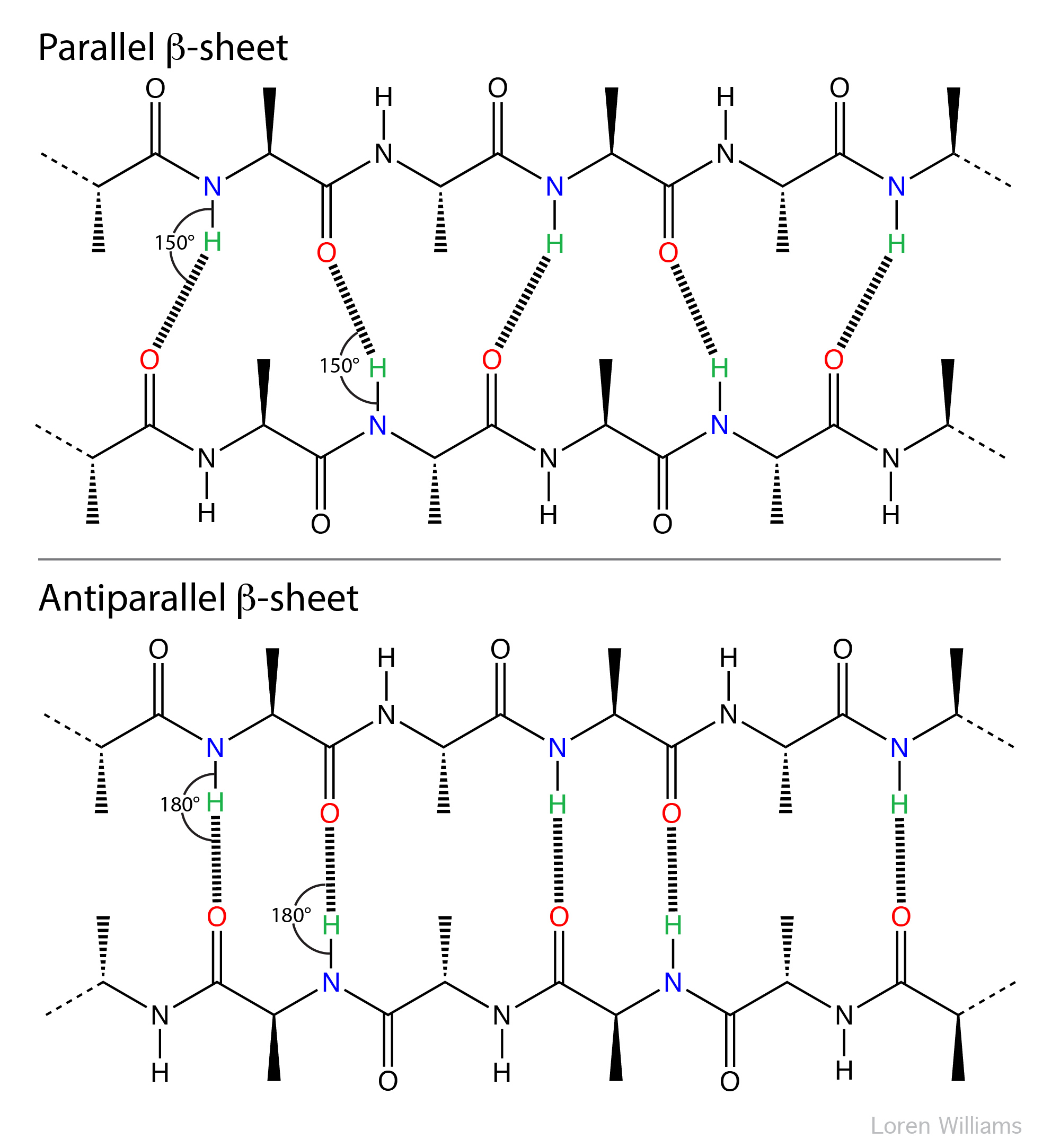
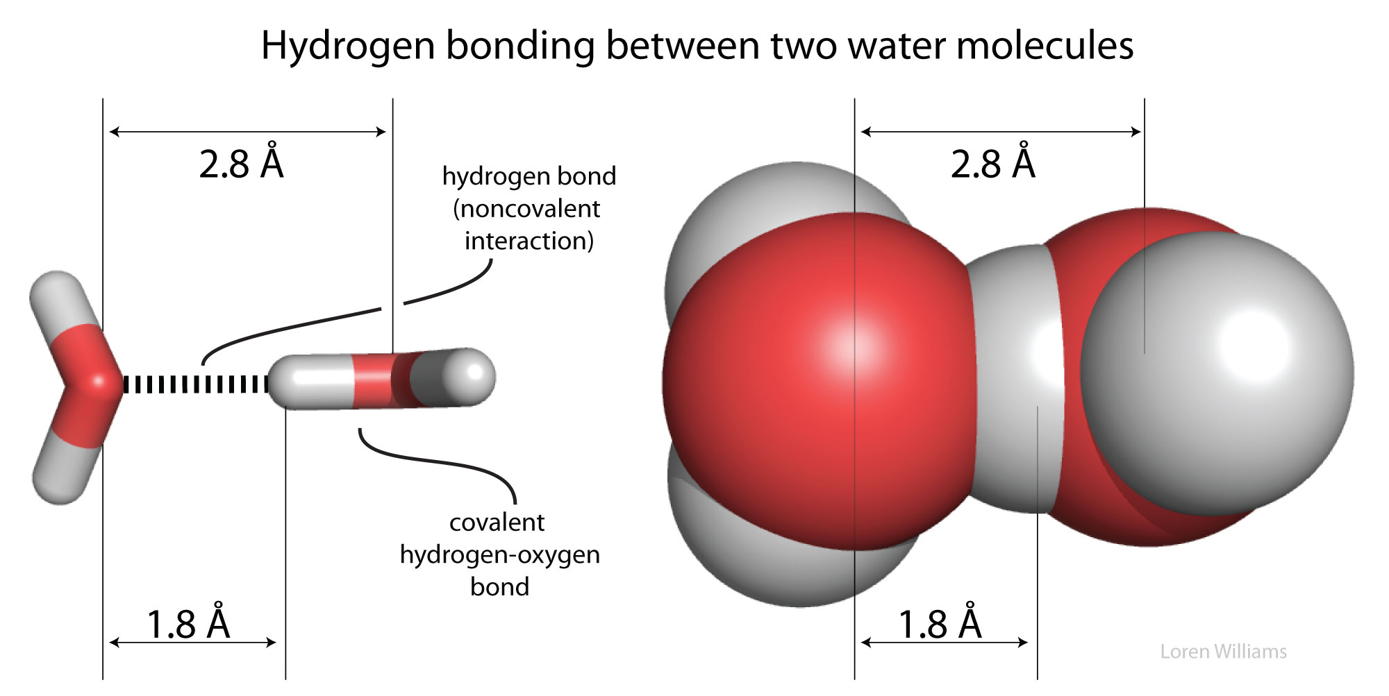
The geometry of a hydrogen bond is defined by three parameters: the covalent donor distance (D–H), the non-covalent acceptor distance (H···A), and the bond angle (∠D–H···A). For an optimal O–H···O interaction, the covalent O–H distance is ∼1.0 Å and the non-covalent H···O distance is ∼1.8 Å (yielding a total heavy-atom O···O separation of ∼2.8 Å). Hydrogen bonds are energetically optimal when nearly linear (∠D–H···A ≈ 180°). In protein secondary structure, this directional requirement is evident in β-sheets: antiparallel β-sheets feature nearly ideal, linear interstrand hydrogen bonds, whereas parallel β-sheets feature bent hydrogen bonds.
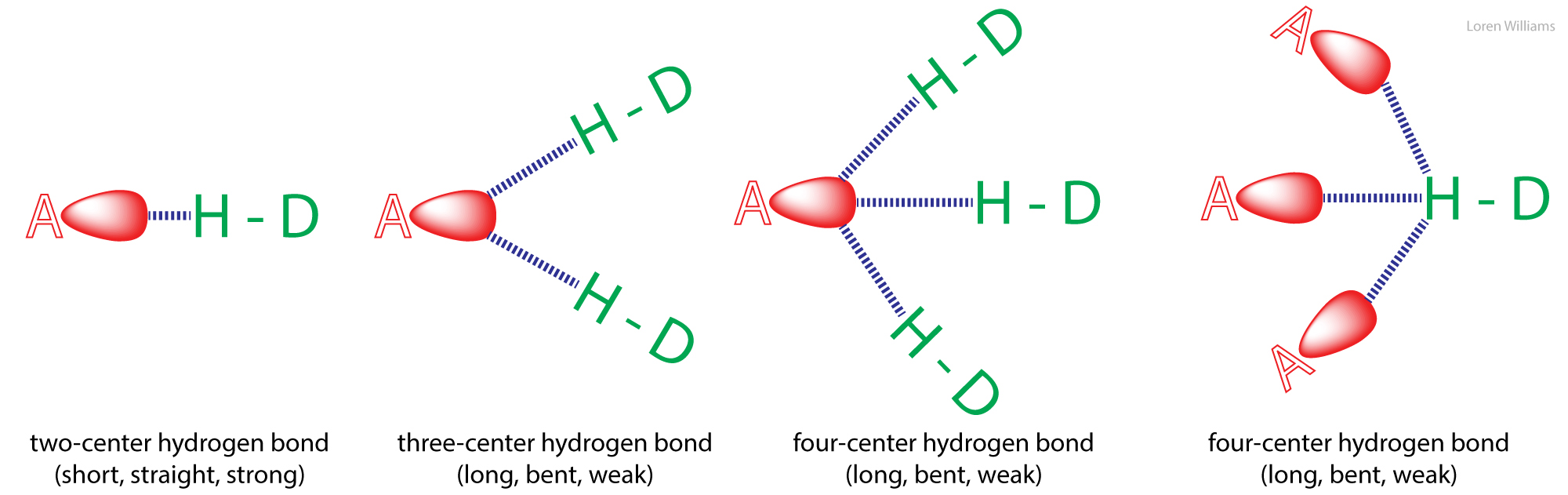
Hydrogen bonds can be two-center (as in β-sheets and ideal ice), three-center, or four-center. Two-center hydrogen bonds are generally shorter, more linear, and stronger than three- or four-center bonds. Three-center hydrogen bonds are also called bifurcated hydrogen bonds, while four-center hydrogen bonds are called trifurcated hydrogen bonds.
Hydrogen atoms are not observable by X-ray crystallography as applied to proteins and nucleic acids, and most PDB entries lack them. Therefore, a geometric description based on hydrogen position is not always practical. In these cases, analysis is usually limited to the D-to-A distance. A hydrogen bond is commonly assigned if the distance between D and A is less than the sum of their van der Waals radii. However, this limit is likely too conservative; an accepted criterion is a distance under 3.4 Å between D and A.
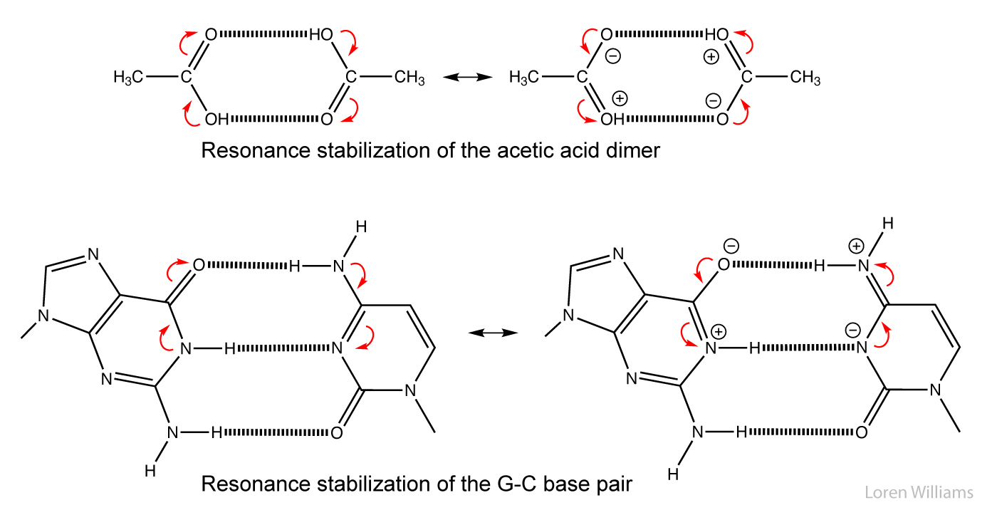
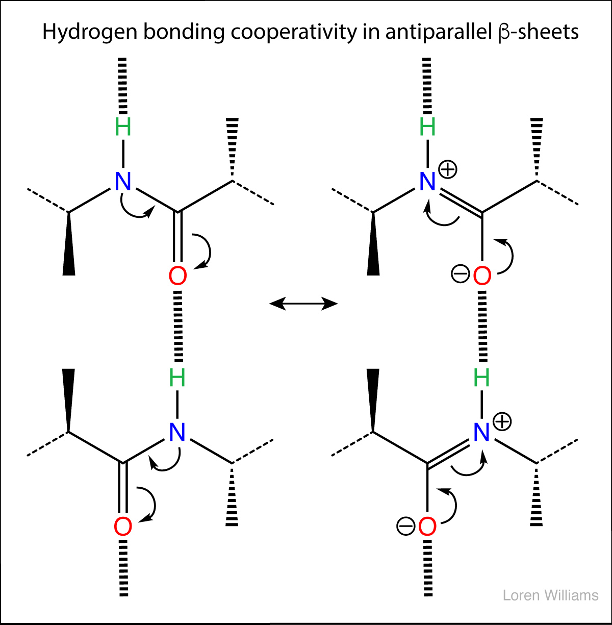
In biological systems, hydrogen bonds are frequently cooperative and stabilized by resonance across multiple bonds. The strength of one hydrogen bond increases that of an adjacent bond. For example, in the acetic acid dimer below, the top bond increases both the acidity of the hydrogen and the basicity of the oxygen in the bottom bond, making each stronger than in isolation. Hydrogen-bond cooperativity occurs widely in base pairing and folded proteins.
H. Water - the liquid of life.
You are not a drop in the ocean. You are the entire ocean in a drop. — Rumi
A drop of water, if it could write out its own history, would explain the universe to us. — Lucy Larcom
Water is the most perfect traveler because when it travels it becomes the path itself. — Mehmet Murat Ildan
Water is the vehicle of nature. — Leonardo da Vinci
Water is not necessary to life; water is life itself. — Antoine de Saint-Exupéry
Water, the most abundant molecule on Earth's surface and the third most abundant in the universe after H2 and CO, is the medium of biology. Most cells are around 65% water by volume and 70% by mass. Organisms have evolved sophisticated mechanisms to acquire and conserve water.
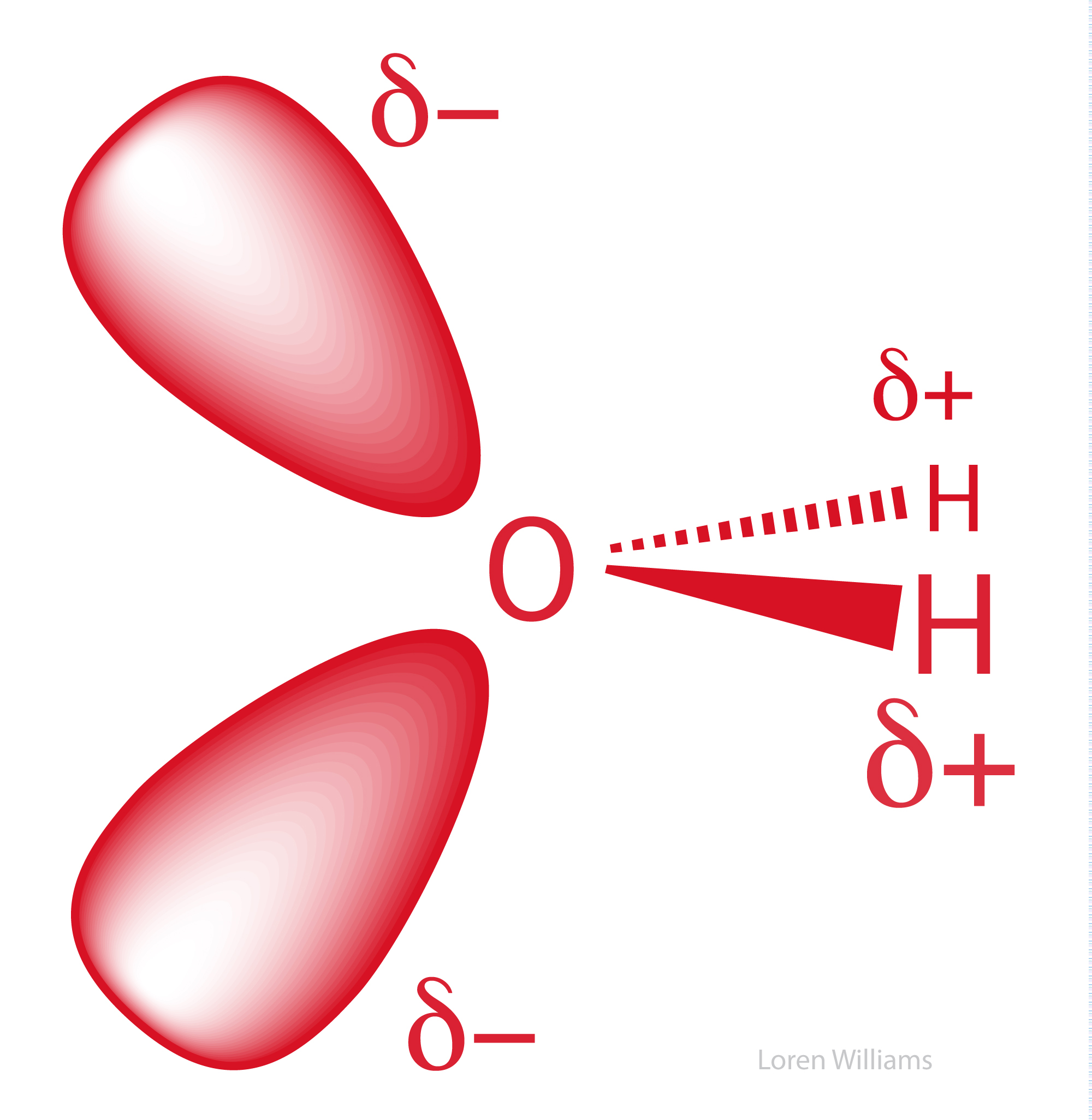
Water is an effective solvent for ions and polar substances, but a poor solvent for nonpolar substances. It drives amphipathic molecules (bearing both polar and nonpolar groups) to spontaneously form compartments. In water, membranes assemble and proteins fold.
Water effectively shields charged species, strongly attenuating interactions between ions. In solution, the electrostatic force between two ions is inversely proportional to the solvent's dielectric constant. Water’s dielectric constant (80.0) is exceptionally high—over twice that of methanol (33.1) and five times that of ammonia (15.5). By weakening the attractive forces between cations and anions, water dissolves salts.
Water is the most frequent chemical actor in biochemistry. Between a third and a half of known biochemical reactions consume or produce water, and it accounts for over 99% of all metabolites in an E. coli cell by molar concentration. Within a cell, a given water molecule repeatedly serves as a substrate, intermediate, cofactor, and product. Virtually all biological molecules are substrates or products of reactions that chemically transform water. Water is never isolated from biological macromolecules, organic cofactors, and metal ions, but readily combines with, dissociates from, and mediates their transformations. It is fully integrated into bond-making and bond-breaking: for biological water, there is no meaningful distinction between medium and chemical participant.
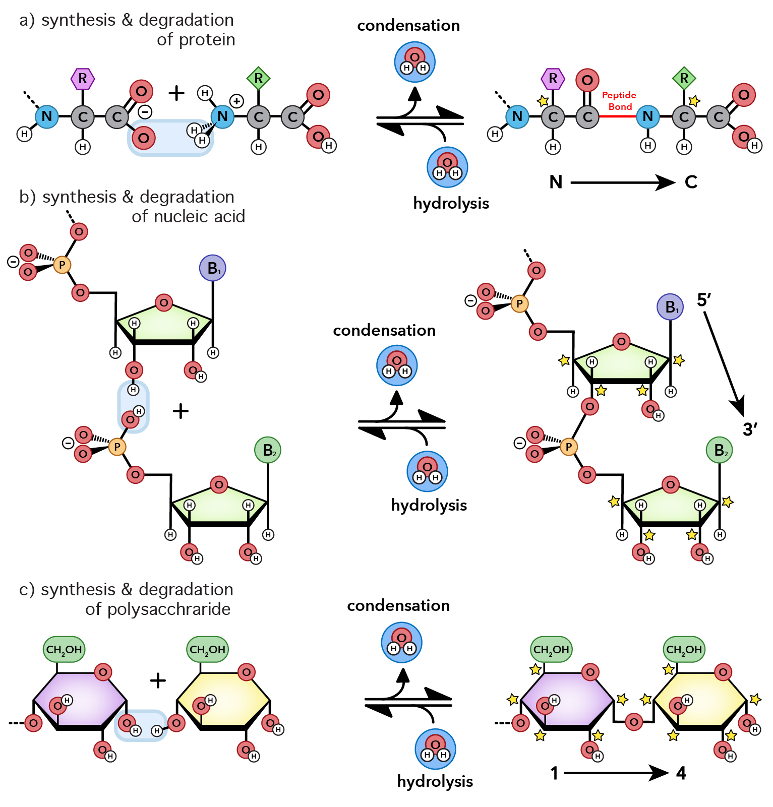
Water acts as both reactant and product in biopolymer synthesis and degradation. All biopolymers form via condensation reactions that eliminate water to link building blocks. For example, joining two amino acids to form a peptide bond yields one water molecule. Conversely, biopolymers degrade via hydrolysis, which consumes water to break these bonds.
Polynucleotides (DNA and RNA) form through the condensation of nucleotides, which are themselves built from smaller subunits by condensation. Similarly, triglycerides and phospholipids form by condensing glycerol with fatty acids and other polar groups, while cellulose—the biosphere's most abundant polymer—forms through the condensation of glucose.
In short, water is both the medium of biology and an active participant in its most fundamental reactions. Stay hydrated.
Hydrogen bonds are ubiquitous in organic systems and minerals, but their density in liquid and solid water is unmatched (see table). No other molecule achieves such a high number of cooperative, cohesive interactions per unit volume. Water forms continuous, three-dimensional hydrogen-bond networks whose density and dynamic rearrangement mediate chemical interactions in ways no other substance can. These dense, geometrically optimized, dynamic, and cooperative networks underlie water’s unusual dielectric constant, density, heat capacity, pH buffering capacity, nucleophile and electrophile activation, and the hydrophobic effect.
| Liquid | Unique H-bonds/molecule |
H-bond/mL (×1022) |
|---|---|---|
| Water | 2.0 | 6.68 |
| Hydrogen Fluoride | 1.1 | 3.28 |
| Formamide | 1.7 | 2.65 |
| Ammonia | 0.75 | 1.81 |
| Hydrogen Sulfide | 0.8 | 1.28 |
| Formic Acid | 1.0 | 1.60 |
| Glycerol | 1.75 | 1.47 |
| Methanol | 0.85 | 1.27 |
Water has an equal number of hydrogen-bond donors and acceptors (two of each). In condensed phases, each molecule can donate two hydrogen bonds and accept two in a tetrahedral geometry. This self-complementarity is an emergent feature of the bulk condensed phase; isolated molecules and small clusters cannot satisfy all four interactions simultaneously.
These strong self-complementary interactions give water exceptionally high melting and boiling points, heats of vaporization and fusion, and surface tension. Water expands upon freezing, causing ice to float. Its heat of vaporization (540 cal/g) is more than twice that of methanol (263 cal/g) and nearly ten times that of chloroform (59 cal/g).
A water molecule (H2O) can form strong hydrogen bonds, with either hydrogen bond donors or acceptors.
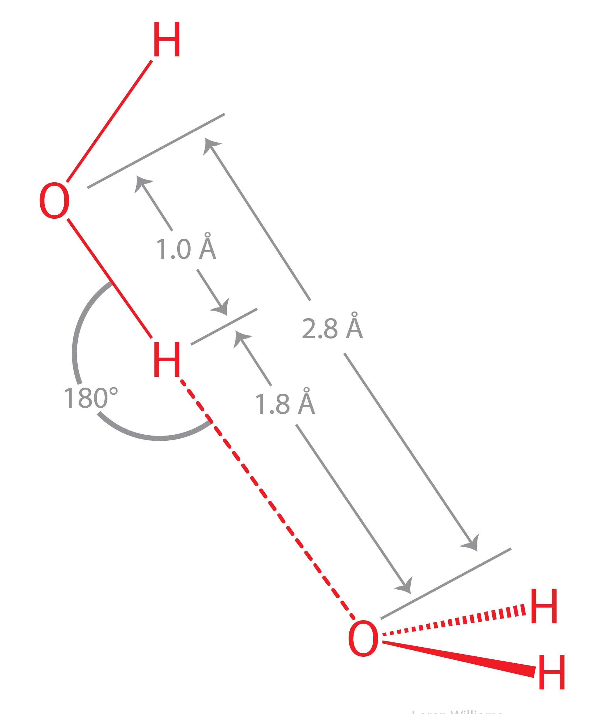
Hydrogen bonds draw atoms closer than their standard van der Waals contact distances. The noncovalent H-to-O distance (~1.8 Å) is shorter than the sum of their van der Waals radii (rH + rO = 1.0 Å + 1.5 Å = 2.5 Å). Similarly, the O-to-O distance (~2.8 Å) is less than twice oxygen’s van der Waals radius (2 × 1.5 Å = 3.0 Å).
The oxygen of a water molecule has four filled valence orbitals (sp3 hybridized) that form a modestly distorted tetrahedron. Two of the electron pairs form covalent bonds with hydrogen atoms and two are non-bonding. The non-bonding lone pairs take more space than the bonding lone pairs, causing the distortion from a perfect tetrahedron. It is useful to imagine that a water molecule is a tetrahedron with negative charge on two apexes and positive charge on two apexes.
X-ray and neutron diffraction of crystalline ice shows that each water molecule is engaged in four hydrogen bonds with intermolecular oxygen-oxygen distances of 2.76 Å. Each oxygen atom is located at the center of a tetrahedron formed by four other oxygen atoms. Each hydrogen atom lies on a line between two oxygen atoms and forms a covalent bond to one oxygen (bond length: 1.00 Å) and a hydrogen bond to the other (hydrogen bond length: 1.76 Å). The tetrahedral shape of an individual water molecule is projected out into the surrounding crystal lattice.
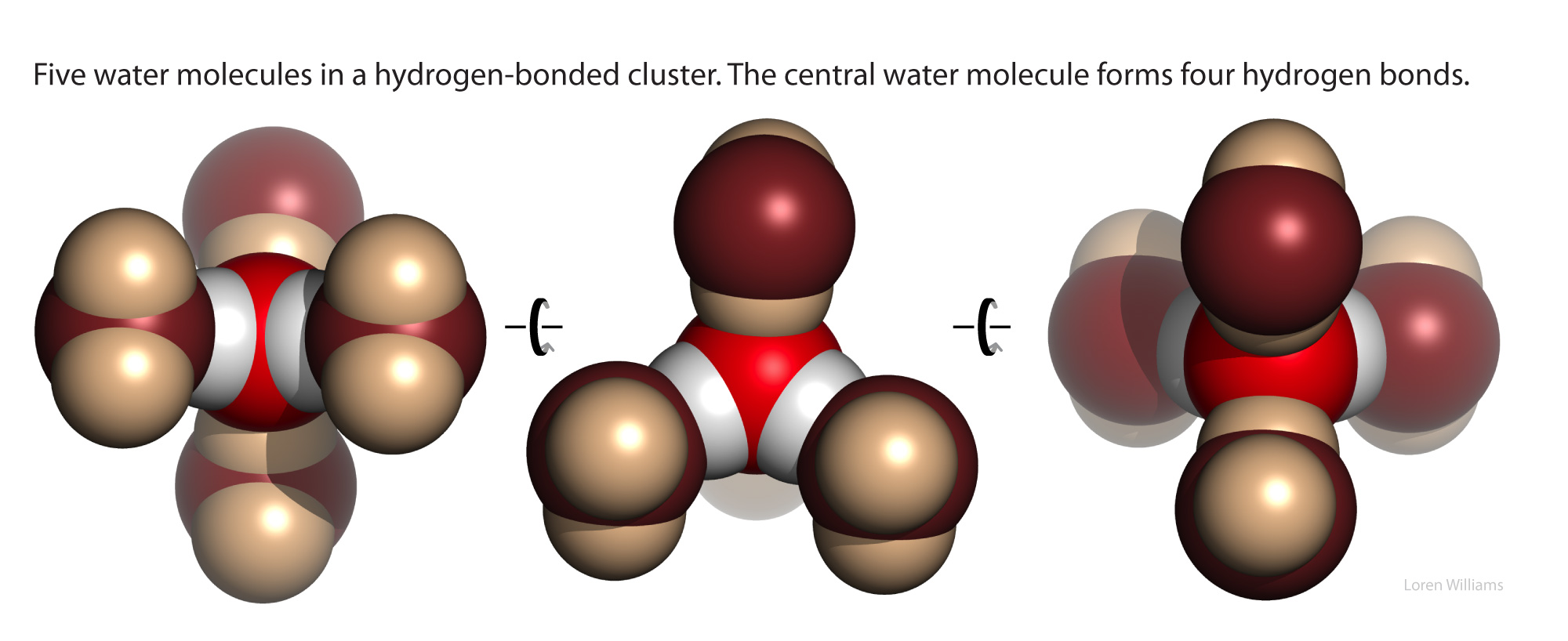
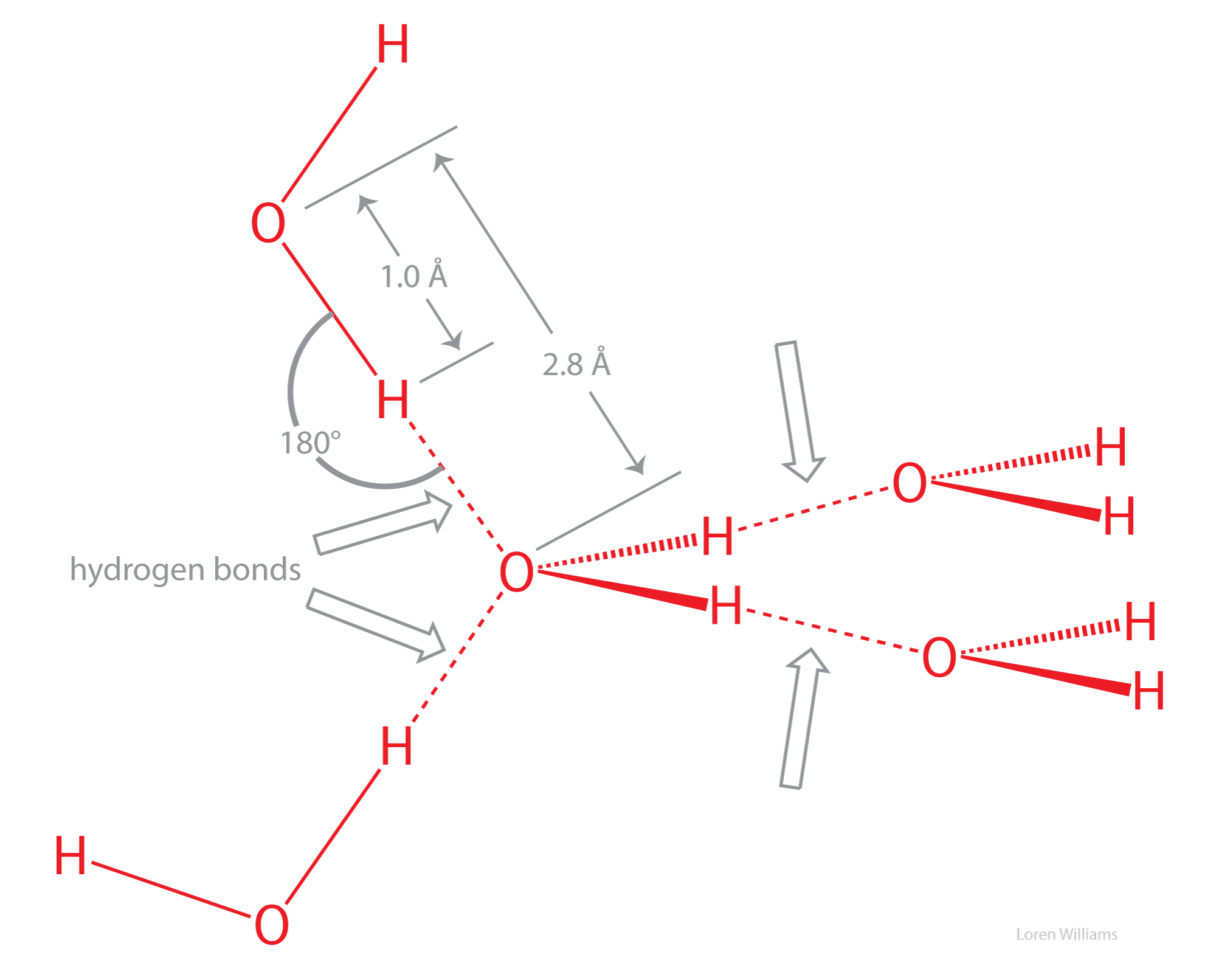
In the crystalline state, directional hydrogen bonding dominates molecular packing, creating open cavities within the lattice. The presence of these cavities in the solid—but not in the liquid—explains why water expands upon freezing. Within the crystal, multiple hydrogen-bond donor–acceptor configurations interconvert through cooperative rotations, allowing water molecules to rotate readily in ice.
Upon melting, thermal energy partially disrupts the rigid, open lattice of ice. Liquid water retains a dynamic, fluctuating network of hydrogen bonds, but the breakdown of the crystalline framework allows molecules to slip into the previously open cavities. This closer interstitial packing increases the number of molecules per unit volume, making liquid water denser than ice.
Comparing ammonia (NH3) with water (H2O) illustrates the importance of water’s self-complementarity. Whereas water features a balanced match of two hydrogen-bond donors and two acceptors, ammonia is noncomplementary, possessing three donors (N–H groups) but only a single lone-pair acceptor.
An isolated ammonia molecule, just like a water molecule, can form strong hydrogen bonds with either hydrogen bond donors or acceptors. Ammonia is more basic than water, and therefore ammonia is a better hydrogen bond acceptor than water.
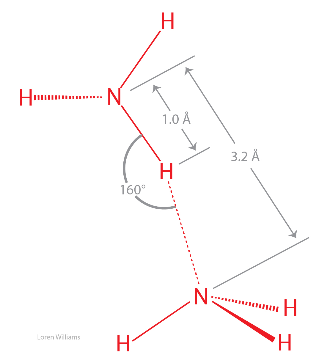
In the crystalline and liquid states, the lone pair of electrons on each nitrogen is shared by multiple hydrogen bond donors. The hydrogen bonds are bifurcated and trifurcated, as described above. The hydrogen bonds in crystalline and liquid ammonia are long, bent and weak.
The boiling point of ammonia is −33 °C, much lower than that of water (100 °C), indicating that molecular interactions in NH3(liq) are significantly weaker than in H2O(liq). The coordinates of an ammonia molecule are here [coordinates].
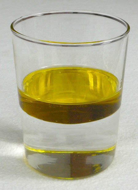
Mixing is generally spontaneous. Whether combining water and ethanol, N2(g) and O2(g), or red and blue marbles, mixing increases entropy by expanding the number of accessible states: there are vastly more ways for components to be mixed than unmixed.
Yet if olive oil and water are mixed by vigorous shaking, they spontaneously unmix. This counterintuitive separation is the hydrophobic effect in action: the exclusion of nonpolar substances from water.
The spontaneous unmixing of olive oil and water is driven by water rather than attractive forces between oil molecules; water actively expels the oil, which is a passive participant. Olive oil molecules self-interact primarily through dispersion forces, whereas water interacts with oil via both dispersion and dipole–induced dipole interactions. In fact, the attractive forces between oil and water molecules are slightly stronger than those within pure oil.
The hydrophobic effect can be understood only through water itself. The phenomenon is entirely a property of the solvent—an indirect consequence of water’s distinctive molecular structure, strong and complementary directional interactions, and unique cohesive properties.
Nomenclature. A hydrophobic molecule is nonpolar, cannot form hydrogen bonds, and dissolves in nonpolar solvents (such as CCl4, cyclohexane, or hydrocarbons) rather than water. A hydrophilic molecule, such as glucose, is polar, forms hydrogen bonds, and is water-soluble. (Cellulose is also polar and hydrophilic, but insoluble in water due to strong interchain cohesion.) An amphipath contains distinct hydrophilic and hydrophobic regions, enabling molecules like phospholipids to assemble into bilayers and micelles. A hydrotrope, such as ATP, is a small amphiphile that solubilizes hydrophobic compounds and prevents aggregation, but lacks the nonpolar volume required to assemble into micelles or bilayers.
We can understand the hydrophobic effect in two separate steps - first a molecular step, then a thermodynamic step.
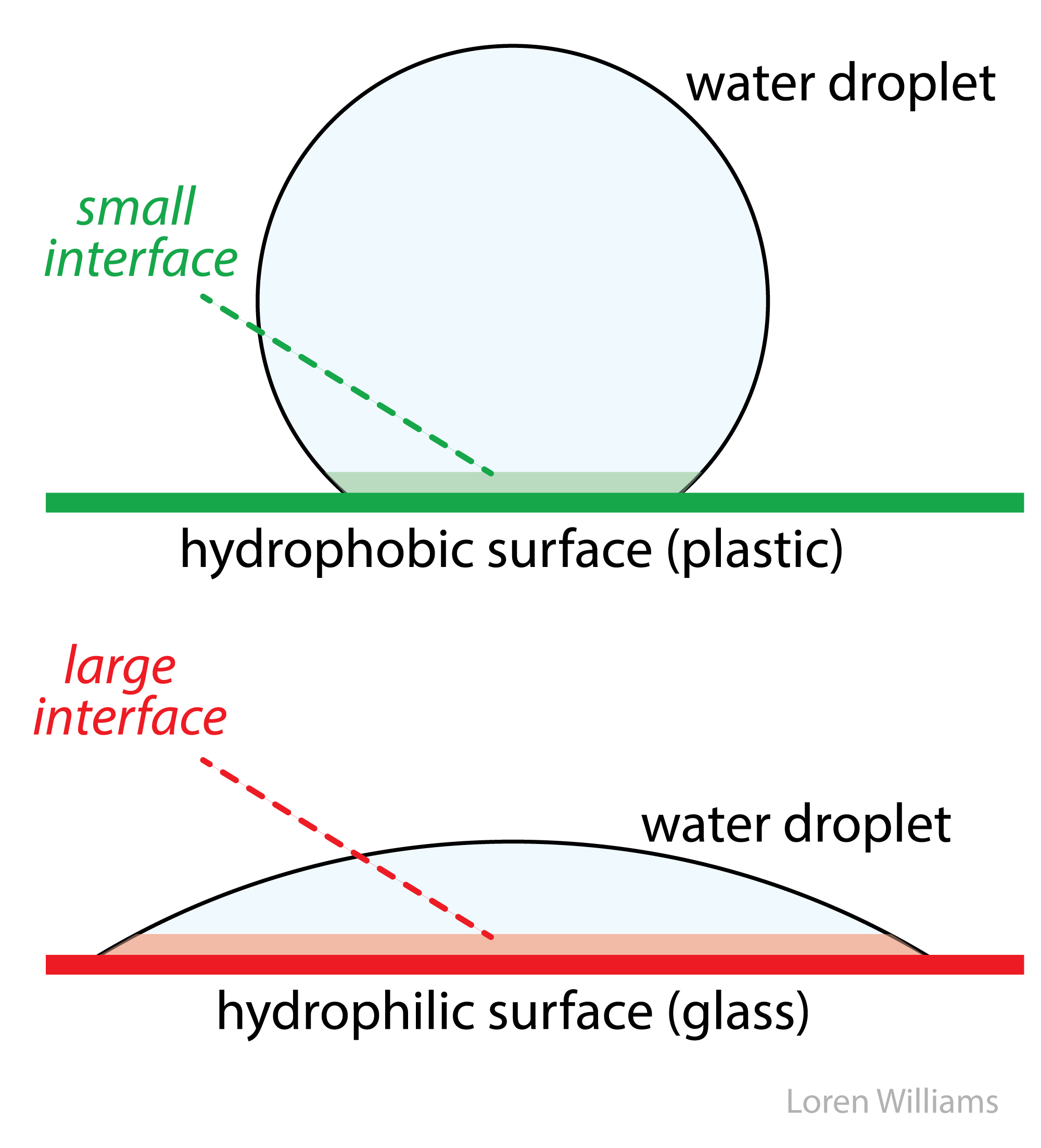
Water–water hydrogen bonding predominates under virtually all conditions. Even when forced into contact with a nonpolar substance or surface, water maintains its hydrogen-bond network—at the cost of distorted geometries and restricted rotational and translational freedom. This ordered interfacial water is thermodynamically unfavorable due to its low entropy. The system maximizes entropy and stability by minimizing interfacial surface area: droplets bead to reduce contact with hydrophobic surfaces, and bulk water actively expels nonpolar solutes from solution.
In bulk water, intermolecular forces are effectively isotropic; molecules can rotate and exchange partners while maintaining a full hydrogen-bond network. At a hydrophobic interface, however, this symmetry is broken. Because the nonpolar substance cannot form hydrogen bonds, local interactions become anisotropic, restraining water’s rotational and translational freedom. Although dispersing the solute increases configurational entropy, the solvent’s loss of orientational entropy heavily dominates, resulting in a net negative entropy of mixing (ΔS < 0) and an unfavorable free energy (ΔG = ΔH − TΔS > 0).
Our description of the hydrophobic effect as an entropic phenomenon is correct only at low (biological) temperatures. We stay in this realm because biochemists don't have to worry about high temperatures. And the term 'hydrophobic bond' is a misnomer and should be avoided, even though Walter Kauzmann, the discoverer of the hydrophobic effect, did often use that phrase.
The molecular descriptions of the hydrophobic effect above can be understood by the thermodynamic parameters enthalpy (ΔH, indicates changes in molecular interactions) and entropy (ΔS, indicates changes in available rotational, translational, vibrational states, etc). A hydrocarbon engages in favorable molecular interactions with water in aqueous solution. We know this because the transfer of a mole of hydrocarbon from pure hydrocarbon to dilute aqueous solution has an enthalpy of around zero. So why don't oil and water mix? It is the water. Water drives non-polar substances out of the aqueous phase.
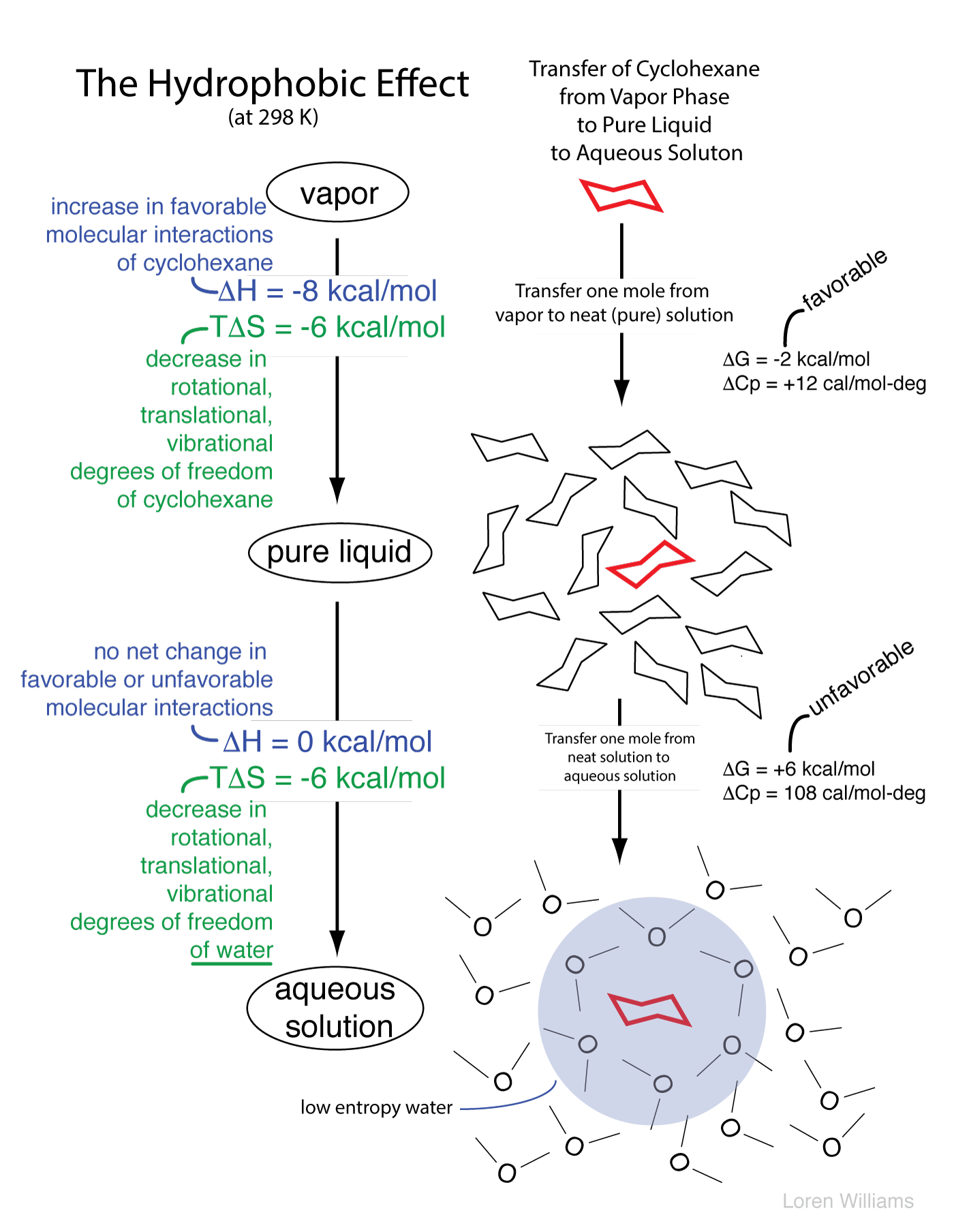
As illustrated below, in the aqueous phase a region of relatively low entropy (high order) water forms at the interface between the aqueous solvent and a hydrophobic solute.
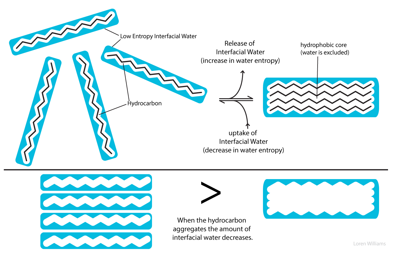
When hydrocarbon molecules aggregate in aqueous solution, the total volume of interfacial water decreases. Thus the driving force for aggregation of hydrophobic substances arises from an increase in entropy of the water. The driving force for aggregation does not arise from intrinsic attraction between hydrophobic solute molecules.
If one considers the entropy of the hydrocarbon molecules alone, a dispersed solution has greater entropy, and is more stable, than an aggregated state. Similarly, a protein may appear to have greater entropy in a random coil than in a native state. Only when the entropy of the aqueous phase is factored into the equation can one understand the separation of water and oil into two phases, and the folding of a protein into a native state.
I. Counterion release.
For many purposes it is useful to approximate DNA as a rod coated with anionic charge. In aqueous solution the negative rod is surrounded by counterions (cations such as Na+, K+ and Mg2+ and/or by polyamines). Counterion release explains much of the salt dependencies of DNA melting, DNA-protein interactions, RNA folding and DNA condensation.
The high density of negative charge on the rod causes strong radial electric fields. The electric field is strong near the rod and weak far from the rod. These electric fields lead to steep radial gradients of the counterion concentration. The counterion concentration is high near the rod and low far from the rod. The "condensed" counterions are mobile, but are constrained to a small volume near to the DNA.
Theoretical considerations show that the local concentration of a monovalent cation such as K+ near the surface of DNA is around 2 molar. The reasons are not obvious, but the concentration of K+ surrounding DNA is largely independent of the K+ concentration in bulk solution. The electrostatic environment surrounding DNA does not depend on the bulk concentration of counterions.
DNA Melting. When DNA melts, the strands separate. Strand separation releases condensed counterions.
Duplex DNA → strand 1 + strand 2 + free counterions
This relationship explains why the stability of double stranded DNA increases (with higher Tm) as salt concentration (ionic strength) increases. Application of Le Chatelier's principle shows that addition of counterions pushes the equilibrium to the left, toward the duplex.
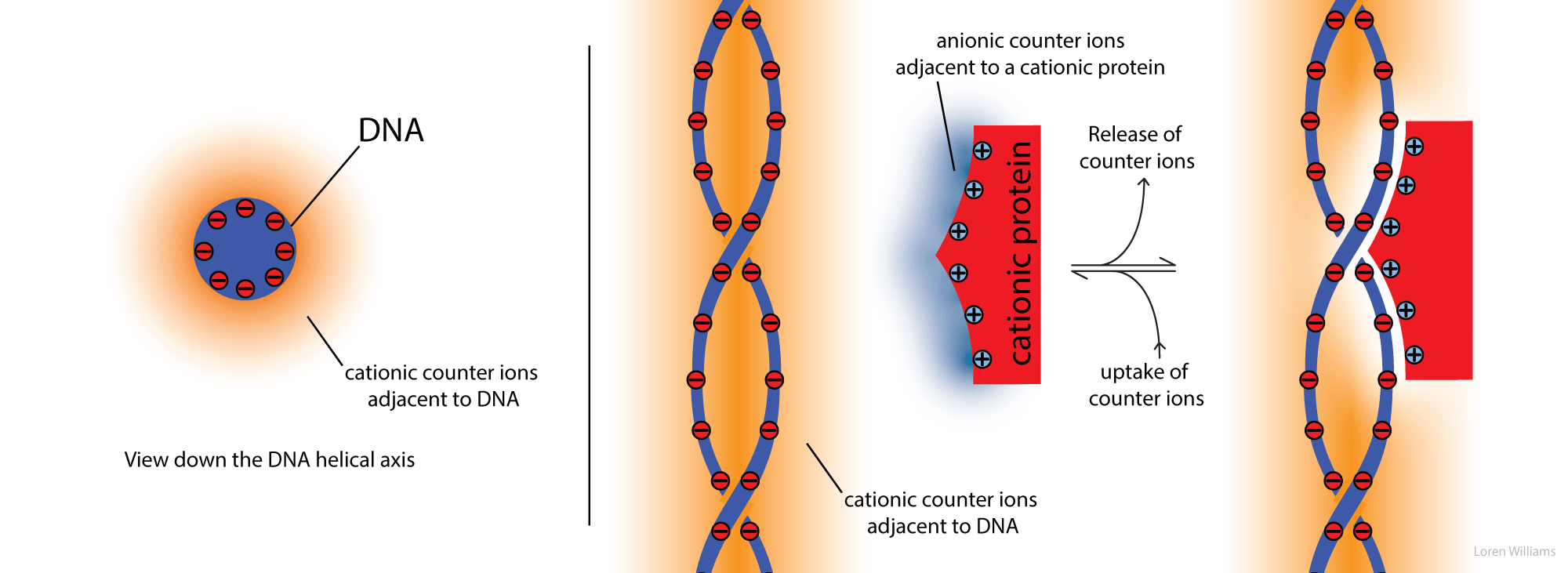
Protein-DNA Interactions. Counterions are released when a cationic protein binds to DNA.
DNA + protein → DNA-protein + free counterions
High salt destabilizes DNA-protein complexes. Cation release explains this salt dependence. Application of Le Chatelier's principle shows that addition of counterions pushes the equilibrium to the left, toward dissociated DNA and dissociated protein.
If the bulk salt concentration is low, there is a large entropic gain from counterion release, and the protein binds tightly to the DNA. If the bulk salt concentration is high, the entropic gain from counterion release is small, and the protein binds weakly.
DNA condensation. Genomic DNAs are very long molecules. The 160,000 base pairs of T4 phage DNA extend to 54 microns. The 4.2 million base pairs of the E. coli chromosome extend to 1.4 millimeters. In biological systems, long DNA molecules must be compacted to fit into very small spaces inside a cell, nucleus or virus particle. The energetic barriers to tight packaging of DNA arise from decreased configurational entropy, bending the stiff double helix, and intermolecular (or inter-segment) electrostatic repulsion of the negatively charged DNA phosphate groups. Yet extended DNA chains condense spontaneously by collapse into very compact, very orderly particles. In the condensed state, DNA helixes are separated by one or two layers of water. Condensed DNA particles are commonly compact toroids. DNA condensation in aqueous solution requires highly charged cations such as spermine (+4) or spermidine (+3). Divalent cations will condense DNA in water-alcohol mixtures. The role of the cations is to decrease electrostatic repulsion of adjacent negatively charged DNA segments. The source of the attraction between nearby DNA segments is not so easy to understand. One possible source of attraction are fluctuations of ion atmospheres in analogy with fluctuating dipoles between molecules (London Forces).
J. About this document.

This document is dedicated to the memory of the late Professor Charles Lochmuller of Duke University. Dr. Lochmuller was a good guy, a natural comic, and an eminent scientist.
Here I approach biochemistry in a new way. It is tradition, starting with Lehninger's first Biochemistry textbook and continuing in essentially all subsequent biochemistry textbooks, to teach about each type of biopolymer in isolation of the others. Protein, DNA, RNA and carbohydrate are described in distinct, well-separated chapters as unrelated chemical phenomena.
In Part 2 of this document I present DNA, RNA, polypeptide, and polysaccharide in the context of their common attributes. Rather than focusing exclusively on the differences (amino acid side chains, nucleic acid bases, etc), I focus on the profound universal properties (self-complementarity, emergence, etc) that unite biopolymers. In my view only by learning about biopolymers in context of each other can one hope to achieve a reasonable understanding of them.
I was fortunate to learn molecular interactions from Dr. Lochmuller in his separations class. I have extended Dr. Lochmuller's parsing scheme to include cation-Π, etc. Some of the source material for Part 1 of document is my 1984 Ph.D. thesis. I wrote core elements in around 1990-92, and expand, revise and clarify the figures and text when inspiration strikes and time is available. The document has benefitted from many discussions with Professor Nicholas Hud.
I created this resource and continue to improve it based on a few core convictions. Molecular interactions are a key to understanding essentially all biological structures, reactions, and processes, and they are best understood using clear illustrations of 3D structures. Because treatments of molecular interactions in textbooks and elsewhere on the web are frequently incomplete, incorrect, or incoherent, all types of molecular interactions can and should be described in a unified and consistent format and style. Furthermore, the molecular interactions of biopolymers are emergent on polymerization. Finally, students around the world from a variety of disciplines are seeking information on molecular interactions, sometimes to answer a specific question (such as what is London Dispersion?) and sometimes seeking more global and systematic information.
The development of this document has been supported by the NASA Astrobiology Institute, the National Science Foundation and the School of Chemistry and Biochemistry at Georgia Tech, all of whom have supported my research laboratory and my public outreach efforts. Comments and suggestions for improvements are welcome and should be addressed to ldw@gatech.edu. I am hopeful that students, especially those who lack resources for textbooks, find this site to be useful.
Reuse. The images and text here can be reused with attribution for noncommercial purposes.
Sincerely,
Loren Williams,
Professor
Georgia Tech
